• 文献检索
  • 文档翻译
  • 深度研究
  • 学术资讯
  • Suppr Zotero 插件Zotero 插件
  • 邀请有礼
  • 套餐&价格
  • 历史记录
应用&插件
Suppr Zotero 插件Zotero 插件浏览器插件Mac 客户端Windows 客户端微信小程序
定价
高级版会员购买积分包购买API积分包
服务
文献检索文档翻译深度研究API 文档MCP 服务
关于我们
关于 Suppr公司介绍联系我们用户协议隐私条款
关注我们

Suppr 超能文献

核心技术专利:CN118964589B侵权必究
粤ICP备2023148730 号-1Suppr @ 2026

文献检索

告别复杂PubMed语法,用中文像聊天一样搜索,搜遍4000万医学文献。AI智能推荐,让科研检索更轻松。

立即免费搜索

文件翻译

保留排版,准确专业,支持PDF/Word/PPT等文件格式,支持 12+语言互译。

免费翻译文档

深度研究

AI帮你快速写综述,25分钟生成高质量综述,智能提取关键信息,辅助科研写作。

立即免费体验

原发性肝神经内分泌癌的影像学及病理特征:9例分析并文献复习

Imaging and pathological features of primary hepatic neuroendocrine carcinoma: An analysis of nine cases and review of the literature.

作者信息

Chen Zhu, Xiao Hua-En, Ramchandra Paudel, Huang Hai-Jiang

机构信息

Department of Radiology, The Second Xiangya Hospital of Central South University, Changsha, Hunan 410011, P.R. China.

Department of Pathology, The Second Xiangya Hospital of Central South University, Changsha, Hunan 410011, P.R. China.

出版信息

Oncol Lett. 2014 Apr;7(4):956-962. doi: 10.3892/ol.2014.1844. Epub 2014 Jan 31.

DOI:10.3892/ol.2014.1844
PMID:24944650
原文链接:https://pmc.ncbi.nlm.nih.gov/articles/PMC3961293/
Abstract

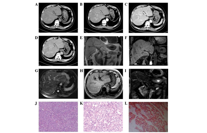
The present study aimed to analyze the imaging features and pathological basis of primary hepatic neuroendocrine carcinoma (PHNEC). A retrospective analysis of the imaging and pathological features of nine PHNEC cases was carried out at The Second Xiangya Hospital of Central South University (Changsha, China). The nine patients were subjected to dynamic contrast-enhanced computed tomography (CT) scanning of the liver and pathological diagnosis of the tissue samples. In addition, two patients were subjected to magnetic resonance imaging (MRI). CT scanning revealed the presence of single or multiple masses in the liver with a maximum diameter of 1-10 cm. These hepatic masses were of low density as showed by plain CT. These masses showed uneven or annular enhancement at their margins in the arterial phase. The venous portal phase showed consistent or declined enhancement and the delayed phase showed light enhancement in these masses. In addition, multiple intrahepatic nodules with long T1 and T2 signal intensities and obvious enhancement were observed by MRI in one patient, while intrahepatic lesions with moderate length T2 signal intensities and light enhancement not visible on the T1 image were observed in another patient. Pathological analysis revealed that the tumor cells exhibited morphological diversity. Immunohistochemical staining revealed that the tumor cells were chromogranin A- and synaptophysin-positive, and carcinoembryonic antigen-, hepatocytic antigen- and α-fetoprotein-negative. Imaging methods, including CT and MRI, are useful for the diagnosis of PHNEC; however, pathological examination is required for a final, definite diagnosis.

摘要

本研究旨在分析原发性肝神经内分泌癌(PHNEC)的影像学特征及病理基础。对中南大学湘雅二医院(中国长沙)9例PHNEC患者的影像学和病理特征进行回顾性分析。这9例患者均接受了肝脏动态对比增强计算机断层扫描(CT)及组织样本的病理诊断。此外,2例患者还接受了磁共振成像(MRI)检查。CT扫描显示肝脏存在单个或多个肿块,最大直径为1 - 10厘米。平扫CT显示这些肝脏肿块呈低密度。这些肿块在动脉期边缘呈不均匀或环形强化。门静脉期强化一致或减弱,延迟期肿块呈轻度强化。另外,1例患者的MRI检查发现多个肝内结节,T1和T2信号长,强化明显;另1例患者肝内病变T2信号强度中等,T1图像上未见明显强化。病理分析显示肿瘤细胞形态多样。免疫组化染色显示肿瘤细胞嗜铬粒蛋白A和突触素阳性,癌胚抗原、肝细胞抗原和甲胎蛋白阴性。包括CT和MRI在内的影像学方法对PHNEC的诊断有帮助;然而,最终明确诊断仍需病理检查。

https://cdn.ncbi.nlm.nih.gov/pmc/blobs/2eeb/3961293/fc895bdff1c1/OL-07-04-0956-g05.jpg
https://cdn.ncbi.nlm.nih.gov/pmc/blobs/2eeb/3961293/73a2baca286b/OL-07-04-0956-g00.jpg
https://cdn.ncbi.nlm.nih.gov/pmc/blobs/2eeb/3961293/31e17df3743d/OL-07-04-0956-g01.jpg
https://cdn.ncbi.nlm.nih.gov/pmc/blobs/2eeb/3961293/1bc1585ff99f/OL-07-04-0956-g02.jpg
https://cdn.ncbi.nlm.nih.gov/pmc/blobs/2eeb/3961293/4b0a1bb6220b/OL-07-04-0956-g03.jpg
https://cdn.ncbi.nlm.nih.gov/pmc/blobs/2eeb/3961293/5d645c8c441f/OL-07-04-0956-g04.jpg
https://cdn.ncbi.nlm.nih.gov/pmc/blobs/2eeb/3961293/fc895bdff1c1/OL-07-04-0956-g05.jpg

相似文献

1
Imaging and pathological features of primary hepatic neuroendocrine carcinoma: An analysis of nine cases and review of the literature.原发性肝神经内分泌癌的影像学及病理特征:9例分析并文献复习
Oncol Lett. 2014 Apr;7(4):956-962. doi: 10.3892/ol.2014.1844. Epub 2014 Jan 31.
2
[CT and MRI findings of primary hepatic neuroendocrine neoplasm].[原发性肝脏神经内分泌肿瘤的CT与MRI表现]
Zhonghua Zhong Liu Za Zhi. 2017 Aug 23;39(8):600-606. doi: 10.3760/cma.j.issn.0253-3766.2017.08.008.
3
CT and MRI diagnosis of hepatic epithelioid hemangioendothelioma.CT 和 MRI 诊断肝上皮样血管内皮细胞瘤。
Hepatobiliary Pancreat Dis Int. 2010 Apr;9(2):154-8.
4
Clinical and radiological characteristics of Chinese patients with hepatic epithelioid hemangioendothelioma.中国肝上皮样血管内皮瘤患者的临床及影像学特征
Ann Saudi Med. 2013 Jul-Aug;33(4):334-8. doi: 10.5144/0256-4947.2013.334.
5
Radiological and histopathological features of hepatic inflammatory myofibroblastic tumour: analysis of 10 cases.肝炎性肌纤维母细胞瘤的放射学和组织病理学特征:10 例分析。
Clin Radiol. 2013 Nov;68(11):1114-20. doi: 10.1016/j.crad.2013.05.097. Epub 2013 Jul 19.
6
Computer tomography and magnetic resonance image manifestations of primary hepatic neuroendocrine cell carcinomas.原发性肝神经内分泌细胞癌的计算机断层扫描和磁共振成像表现
Asian Pac J Cancer Prev. 2014;15(6):2759-64. doi: 10.7314/apjcp.2014.15.6.2759.
7
Response to hepatic arterial infusion chemotherapy combined with camrelizumab and targeted therapy in advanced primary hepatic neuroendocrine carcinoma: a case report and literature review.肝动脉灌注化疗联合卡瑞利珠单抗及靶向治疗晚期原发性肝神经内分泌癌的疗效:1例病例报告及文献复习
J Gastrointest Oncol. 2024 Aug 31;15(4):1962-1972. doi: 10.21037/jgo-24-571. Epub 2024 Aug 28.
8
Rare hepatic malignant tumors: dynamic CT, MRI, and clinicopathologic features: with analysis of 54 cases and review of the literature.罕见肝脏恶性肿瘤:动态CT、MRI及临床病理特征:附54例分析并文献复习
Abdom Imaging. 2013 Jun;38(3):511-26. doi: 10.1007/s00261-012-9918-y.
9
[Value of computed tomography and magnetic resonance imaging in diagnosis and differential diagnosis of small hepatocellular carcinoma].[计算机断层扫描和磁共振成像在小肝细胞癌诊断及鉴别诊断中的价值]
Nan Fang Yi Ke Da Xue Xue Bao. 2008 Dec;28(12):2235-8.
10
[Diagnostic imaging of primary hepatic neuroendocrine tumors and treatment with transarterial chemoembolization: analysis of 6 cases].[原发性肝脏神经内分泌肿瘤的诊断性影像学及经动脉化疗栓塞治疗:6例分析]
Zhonghua Gan Zang Bing Za Zhi. 2018 Apr 20;26(4):294-297. doi: 10.3760/cma.j.issn.1007-3418.2018.04.012.

引用本文的文献

1
Role of advanced imaging in primary hepatic neuroendocrine tumor with borderline raised AFP and negative chromogranin staining: A case report.高级影像学检查在甲胎蛋白临界升高且嗜铬粒蛋白染色阴性的原发性肝神经内分泌肿瘤中的作用:一例报告
Int J Surg Case Rep. 2024 Dec;125:110647. doi: 10.1016/j.ijscr.2024.110647. Epub 2024 Nov 23.
2
Primary hepatic neuroendocrine neoplasms: imaging characteristics and misdiagnosis analysis.原发性肝脏神经内分泌肿瘤:影像学特征及误诊分析
Front Oncol. 2024 May 14;14:1391663. doi: 10.3389/fonc.2024.1391663. eCollection 2024.
3
Diagnostic dilemma in a patient with history of medullary thyroid carcinoma and abnormal serum liver enzymes; a case report with six years follow up.

本文引用的文献

1
Gastric neuroendocrine tumors in our institutions according to the WHO 2010 classification.根据世界卫生组织2010年分类标准对我们机构中的胃神经内分泌肿瘤进行分类。
Int Surg. 2012 Oct-Dec;97(4):335-9. doi: 10.9738/CC134.1.
2
Primary hepatic neuroendocrine tumour requiring live donor liver transplantation: case report and concise review.原发性肝神经内分泌肿瘤需要活体肝移植:病例报告及简要综述。
Ann Hepatol. 2012 Sep-Oct;11(5):715-20.
3
Primary hepatic neuroendocrine tumor: Five cases with different preoperative diagnoses.原发性肝神经内分泌肿瘤:5例术前诊断不同的病例。
病史为甲状腺髓样癌伴血清肝酶异常患者的诊断困境;一例六年随访病例报告。
BMC Endocr Disord. 2023 Aug 30;23(1):186. doi: 10.1186/s12902-023-01439-7.
4
Comparison of primary hepatic neuroendocrine tumors and non-hepatitis B non-hepatitis C hepatocellular carcinoma on contrast-enhanced ultrasound.原发性肝神经内分泌肿瘤与非乙型非丙型肝炎肝细胞癌的超声造影对比
Front Oncol. 2023 Jul 10;13:1106281. doi: 10.3389/fonc.2023.1106281. eCollection 2023.
5
Primary hepatic neuroendocrine neoplasm presenting as a massive cystic liver tumor mimicking mucinous cystic neoplasm of the liver: A case report and literature review.表现为巨大囊性肝肿瘤并酷似肝黏液性囊性肿瘤的原发性肝神经内分泌肿瘤:1例报告及文献复习
Oncol Lett. 2023 May 31;26(1):304. doi: 10.3892/ol.2023.13890. eCollection 2023 Jul.
6
Primary Hepatic Neuroendocrine Carcinoma with Thrombocytopenia Due to Diffuse Bone Marrow and Splenic Infiltration: An Autopsy Case.原发性肝神经内分泌癌伴血小板减少症,源于骨髓弥漫性浸润和脾脏浸润:尸检病例。
Intern Med. 2022 Nov 15;61(22):3361-3368. doi: 10.2169/internalmedicine.9465-22. Epub 2022 Apr 9.
7
Clinical characteristics and outcome of primary hepatic neuroendocrine tumors after comprehensive therapy.综合治疗后原发性肝脏神经内分泌肿瘤的临床特征及预后
World J Gastrointest Oncol. 2020 Sep 15;12(9):1031-1043. doi: 10.4251/wjgo.v12.i9.1031.
8
Primary hepatic neuroendocrine tumours of liver- a rarity: Single centre analysis of 13 patients.原发性肝脏神经内分泌肿瘤——一种罕见病:13例患者的单中心分析
Ann Hepatobiliary Pancreat Surg. 2020 Feb;24(1):17-23. doi: 10.14701/ahbps.2020.24.1.17. Epub 2020 Feb 27.
9
Primary hepatic neuroendocrine tumors: A case report.原发性肝神经内分泌肿瘤:一例报告。
Medicine (Baltimore). 2019 Dec;98(50):e18278. doi: 10.1097/MD.0000000000018278.
10
Analysis of the clinicopathological features and prognostic factors of primary hepatic neuroendocrine tumors.原发性肝脏神经内分泌肿瘤的临床病理特征及预后因素分析
Oncol Lett. 2018 Jun;15(6):8604-8610. doi: 10.3892/ol.2018.8413. Epub 2018 Apr 2.
Turk J Gastroenterol. 2012 Jun;23(3):272-8. doi: 10.4318/tjg.2012.0465.
4
Primary hepatic neuroendocrine tumor: an unusual cystic presentation.原发性肝神经内分泌肿瘤:一种不寻常的囊性表现。
Indian J Pathol Microbiol. 2010 Oct-Dec;53(4):760-2. doi: 10.4103/0377-4929.72078.
5
Primary hepatic carcinoid: a case report and literature review.原发性肝类癌:一例报告及文献复习
World J Gastroenterol. 2009 May 21;15(19):2418-22. doi: 10.3748/wjg.15.2418.
6
Primary hepatic carcinoid; a diagnostic dilemma: a case report.原发性肝类癌;诊断难题:一例病例报告
Cases J. 2008 Nov 17;1(1):314. doi: 10.1186/1757-1626-1-314.
7
Primary carcinoids of the liver: a review of symptoms, diagnosis and treatments.原发性肝脏类癌:症状、诊断与治疗综述
Dig Surg. 2008;25(5):364-8. doi: 10.1159/000167021. Epub 2008 Nov 4.
8
Primary carcinoid tumors of the liver.原发性肝脏类癌肿瘤。
World J Surg Oncol. 2008 Aug 27;6:91. doi: 10.1186/1477-7819-6-91.
9
Laparoscopic radiofrequency ablation of neuroendocrine liver metastases: a 10-year experience evaluating predictors of survival.腹腔镜射频消融治疗神经内分泌肝转移瘤:一项评估生存预测因素的10年经验
Surgery. 2007 Jul;142(1):10-9. doi: 10.1016/j.surg.2007.01.036.
10
Chromogranin A: is it a useful marker of neuroendocrine tumors?嗜铬粒蛋白A:它是神经内分泌肿瘤的有用标志物吗?
J Clin Oncol. 2007 May 20;25(15):1967-73. doi: 10.1200/JCO.2006.10.1535.