Medical School of Science of Santa Casa de São Paulo (FCMSCSP) / French-Brazilian Centre of Endoscopic Ultrasonography (CFBEUS), Brazil.
Medical School of Science of Santa Casa de São Paulo (FCMSCSP), Brazil.
Endosc Ultrasound. 2012 Apr;1(1):23-35. doi: 10.7178/eus.01.005.
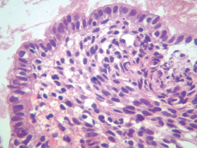
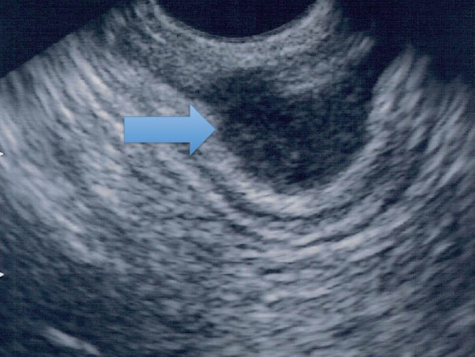
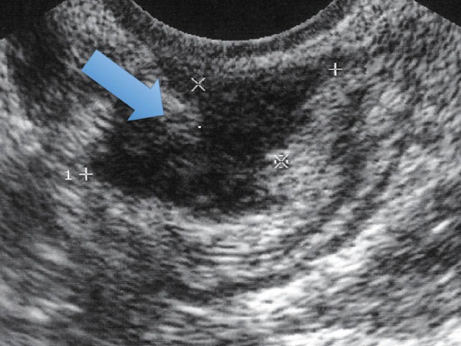
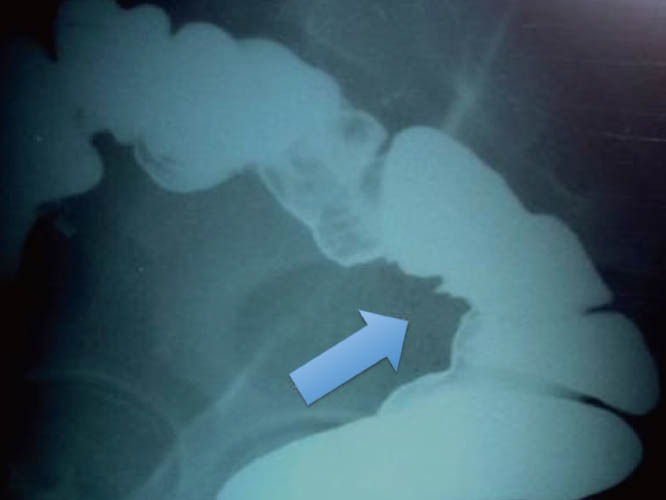
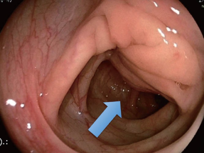
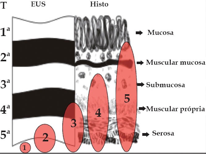
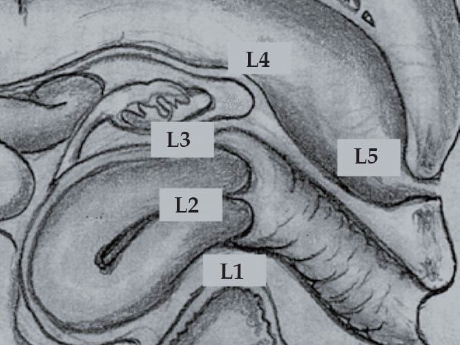
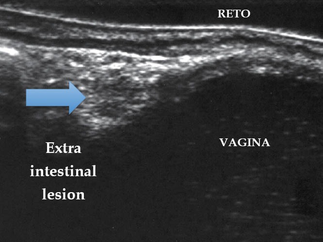
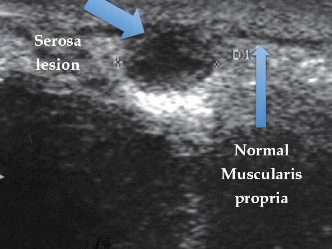
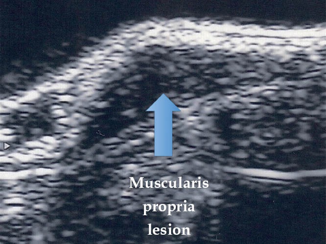
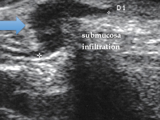
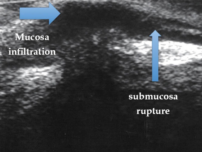
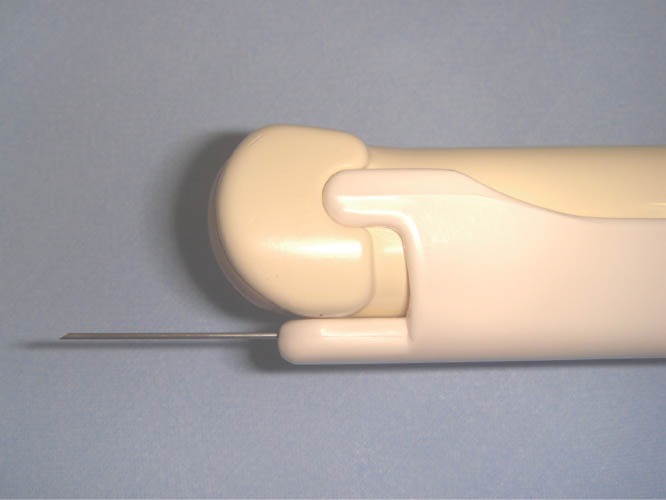
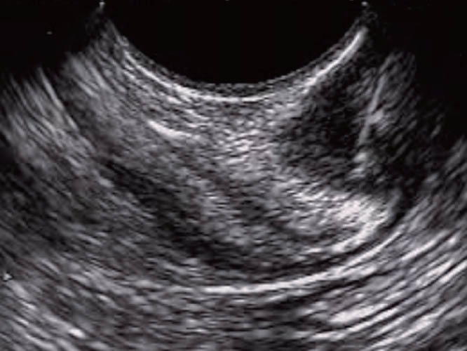
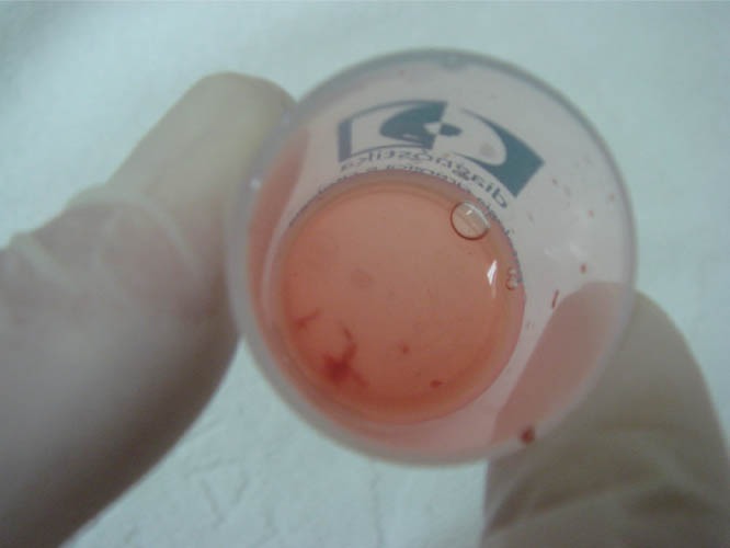
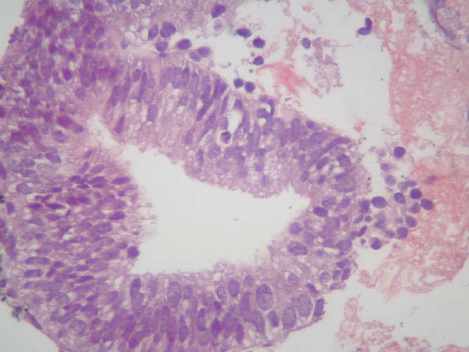
The widespread use of endoscopic ultrasound has facilitated the evaluation of subepithelial and surrounding lesions of the gastrointestinal tract. Deep pelvic endometriosis, with or without infiltration of the intestinal wall, is a frequent disease that can be observed in women in their fertile age. Patients of this disease may present nonspecific signs and symptoms or be completely asymptomatic. Laparoscopic surgical resection of endometriotic lesions is the treatment of choice in symptomatic patients. An accurate preoperative evaluation is indispensable for therapeutic decisions mainly in the suspicion of intestinal wall and/or urinary tract infiltration, and also in cases where we need to establish histological diagnosis or to rule out malignant disease. Diagnostic tools, including transrectal ultrasound, magnetic resonance image, transvaginal ultrasound, barium enema, and colonoscopy, play significant roles in determining the presence, depth, histology, and other relevant data about the extension of the disease. Diagnostic algorithm depends on the clinical presentation, the expertise of the medical team, and the technology available at each institution. This article reviews and discusses relevant clinical points in endometriosis, including techniques and outcomes of the study of the disease through transrectal ultrasound and fine-needle aspiration.
内镜超声的广泛应用促进了胃肠道黏膜下和周围病变的评估。深部盆腔子宫内膜异位症,无论是否累及肠壁,是一种常见疾病,可发生于育龄期妇女。这些患者可能表现为非特异性症状,也可能完全无症状。对于有症状的患者,腹腔镜下切除子宫内膜异位症病灶是治疗的首选方法。准确的术前评估对于治疗决策至关重要,主要是在怀疑肠壁和/或泌尿道浸润,以及需要建立组织学诊断或排除恶性疾病的情况下。诊断工具,包括经直肠超声、磁共振成像、经阴道超声、钡灌肠和结肠镜检查,在确定疾病的存在、深度、组织学和其他与疾病扩展相关的数据方面发挥着重要作用。诊断算法取决于临床表现、医疗团队的专业知识以及每个机构可用的技术。本文回顾和讨论了子宫内膜异位症的相关临床要点,包括经直肠超声和细针抽吸术研究疾病的技术和结果。