• 文献检索
  • 文档翻译
  • 深度研究
  • 学术资讯
  • Suppr Zotero 插件Zotero 插件
  • 邀请有礼
  • 套餐&价格
  • 历史记录
应用&插件
Suppr Zotero 插件Zotero 插件浏览器插件Mac 客户端Windows 客户端微信小程序
定价
高级版会员购买积分包购买API积分包
服务
文献检索文档翻译深度研究API 文档MCP 服务
关于我们
关于 Suppr公司介绍联系我们用户协议隐私条款
关注我们

Suppr 超能文献

核心技术专利:CN118964589B侵权必究
粤ICP备2023148730 号-1Suppr @ 2026

文献检索

告别复杂PubMed语法,用中文像聊天一样搜索,搜遍4000万医学文献。AI智能推荐,让科研检索更轻松。

立即免费搜索

文件翻译

保留排版,准确专业,支持PDF/Word/PPT等文件格式,支持 12+语言互译。

免费翻译文档

深度研究

AI帮你快速写综述,25分钟生成高质量综述,智能提取关键信息,辅助科研写作。

立即免费体验

使用多尺度空间韦伯定律描述符和支持向量机减少乳腺X线摄影中的假阳性

False-positive reduction in mammography using multiscale spatial Weber law descriptor and support vector machines.

作者信息

Hussain Muhammad

机构信息

Department of Software Engineering, College of Computer and Information Sciences, King Saud University, Riyadh, Saudi Arabia.

出版信息

Neural Comput Appl. 2014;25(1):83-93. doi: 10.1007/s00521-013-1450-7. Epub 2013 Jul 13.

DOI:10.1007/s00521-013-1450-7
PMID:24954976
原文链接:https://pmc.ncbi.nlm.nih.gov/articles/PMC4055841/
Abstract

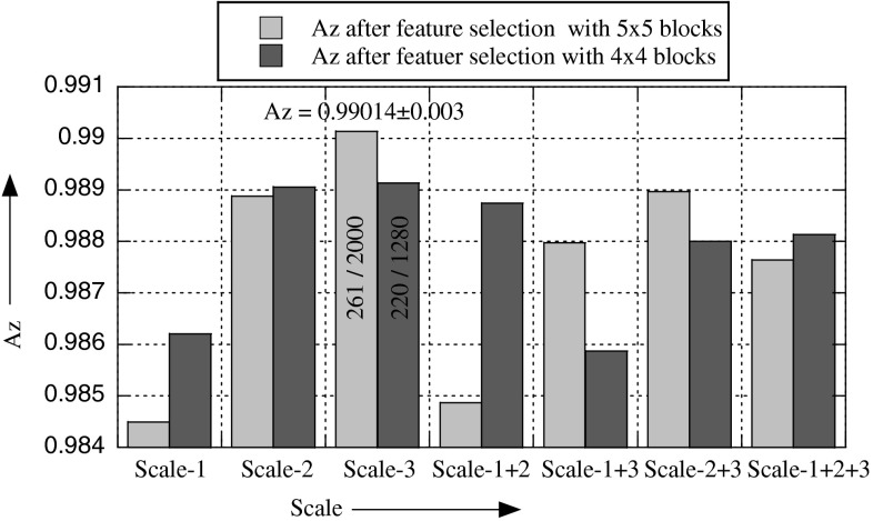
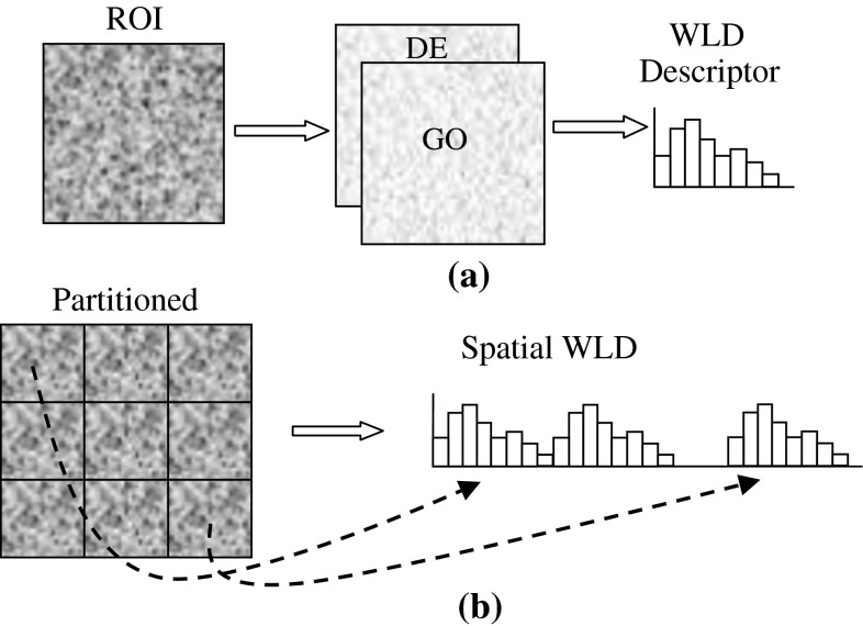
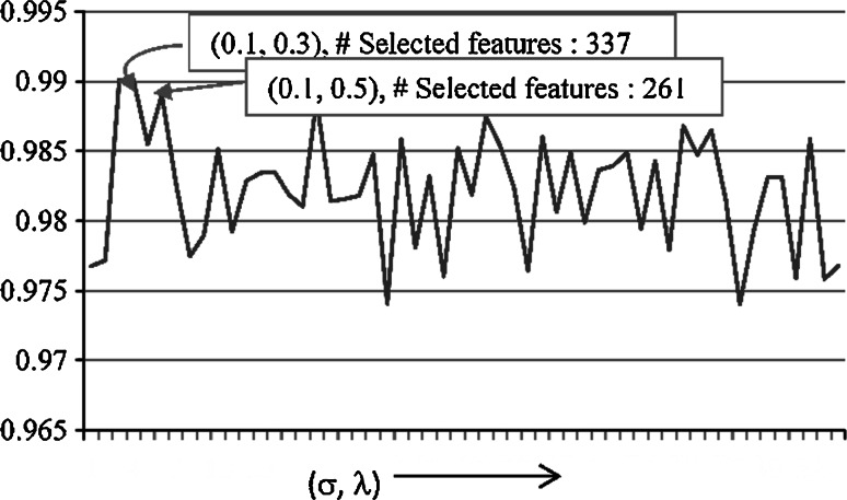
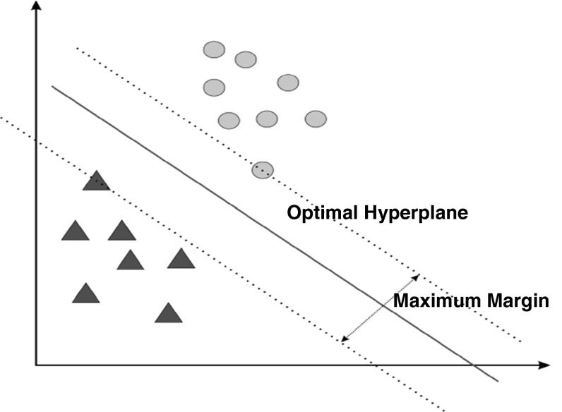
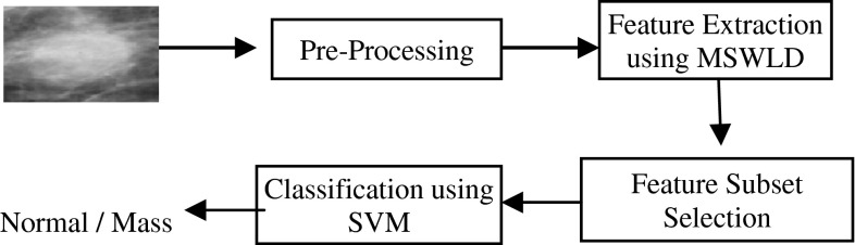
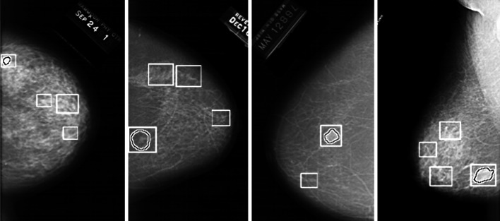
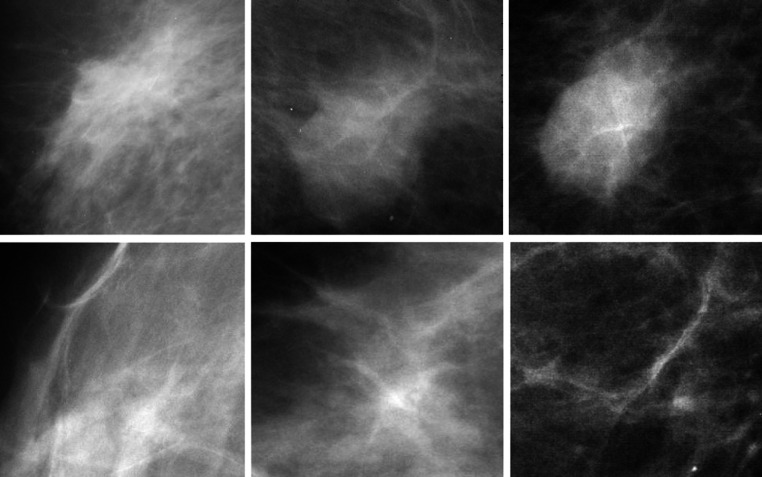
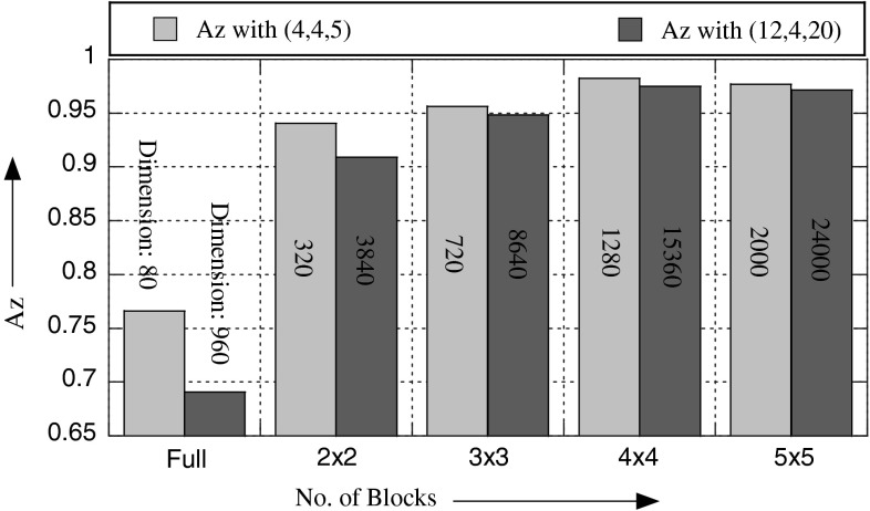
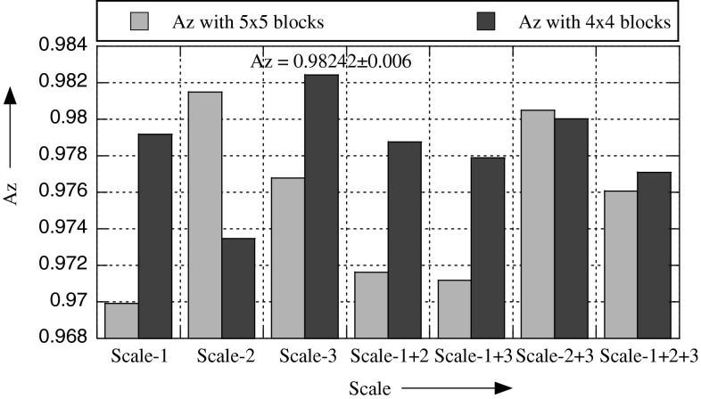
In a CAD system for the detection of masses, segmentation of mammograms yields regions of interest (ROIs), which are not only true masses but also suspicious normal tissues that result in false positives. We introduce a new method for false-positive reduction in this paper. The key idea of our approach is to exploit the textural properties of mammograms and for texture description, to use Weber law descriptor (WLD), which outperforms state-of-the-art best texture descriptors. The basic WLD is a holistic descriptor by its construction because it integrates the local information content into a single histogram, which does not take into account the spatial locality of micropatterns. We extend it into a multiscale spatial WLD (MSWLD) that better characterizes the texture micro structures of masses by incorporating the spatial locality and scale of microstructures. The dimension of the feature space generated by MSWLD becomes high; it is reduced by selecting features based on their significance. Finally, support vector machines are employed to classify ROIs as true masses or normal parenchyma. The proposed approach is evaluated using 1024 ROIs taken from digital database for screening mammography and an accuracy of Az = 0.99 ± 0.003 (area under receiver operating characteristic curve) is obtained. A comparison reveals that the proposed method has significant improvement over the state-of-the-art best methods for false-positive reduction problem.

摘要

在用于肿块检测的CAD系统中,乳腺X光图像的分割会产生感兴趣区域(ROI),这些区域不仅包括真正的肿块,还包括可疑的正常组织,从而导致假阳性。在本文中,我们介绍一种减少假阳性的新方法。我们方法的关键思想是利用乳腺X光图像的纹理特性,并使用韦伯定律描述符(WLD)进行纹理描述,该描述符优于当前最先进的最佳纹理描述符。基本的WLD从其构造来看是一种整体描述符,因为它将局部信息内容整合到单个直方图中,没有考虑微模式的空间局部性。我们将其扩展为多尺度空间WLD(MSWLD),通过纳入微结构的空间局部性和尺度,能更好地表征肿块的纹理微观结构。由MSWLD生成的特征空间维度变高;通过基于特征的重要性进行选择来降低维度。最后,使用支持向量机将ROI分类为真正的肿块或正常实质。使用从乳腺X光筛查数字数据库中获取的1024个ROI对所提出的方法进行评估,得到的Az = 0.99±0.003(接收器操作特征曲线下面积)的准确率。比较结果表明,对于减少假阳性问题,所提出的方法比当前最先进的最佳方法有显著改进。

https://cdn.ncbi.nlm.nih.gov/pmc/blobs/d971/4055841/0bc675e5f723/521_2013_1450_Fig10_HTML.jpg
https://cdn.ncbi.nlm.nih.gov/pmc/blobs/d971/4055841/ca5888415329/521_2013_1450_Fig1_HTML.jpg
https://cdn.ncbi.nlm.nih.gov/pmc/blobs/d971/4055841/dd092d968e75/521_2013_1450_Fig2_HTML.jpg
https://cdn.ncbi.nlm.nih.gov/pmc/blobs/d971/4055841/5ff9bb54a7c8/521_2013_1450_Fig3_HTML.jpg
https://cdn.ncbi.nlm.nih.gov/pmc/blobs/d971/4055841/409648008fd9/521_2013_1450_Fig4_HTML.jpg
https://cdn.ncbi.nlm.nih.gov/pmc/blobs/d971/4055841/d0abae2b6d0f/521_2013_1450_Fig5_HTML.jpg
https://cdn.ncbi.nlm.nih.gov/pmc/blobs/d971/4055841/f8a45565bdab/521_2013_1450_Fig6_HTML.jpg
https://cdn.ncbi.nlm.nih.gov/pmc/blobs/d971/4055841/f9c50e453288/521_2013_1450_Fig7_HTML.jpg
https://cdn.ncbi.nlm.nih.gov/pmc/blobs/d971/4055841/cbda2af4739d/521_2013_1450_Fig8_HTML.jpg
https://cdn.ncbi.nlm.nih.gov/pmc/blobs/d971/4055841/5735354fa5ce/521_2013_1450_Fig9_HTML.jpg
https://cdn.ncbi.nlm.nih.gov/pmc/blobs/d971/4055841/0bc675e5f723/521_2013_1450_Fig10_HTML.jpg

相似文献

1
False-positive reduction in mammography using multiscale spatial Weber law descriptor and support vector machines.使用多尺度空间韦伯定律描述符和支持向量机减少乳腺X线摄影中的假阳性
Neural Comput Appl. 2014;25(1):83-93. doi: 10.1007/s00521-013-1450-7. Epub 2013 Jul 13.
2
A textural approach for mass false positive reduction in mammography.一种用于减少乳腺X线摄影中大量假阳性的纹理分析方法。
Comput Med Imaging Graph. 2009 Sep;33(6):415-22. doi: 10.1016/j.compmedimag.2009.03.007. Epub 2009 Apr 29.
3
False-positive reduction in computer-aided mass detection using mammographic texture analysis and classification.利用乳腺图像纹理分析和分类降低计算机辅助检测的假阳性率。
Comput Methods Programs Biomed. 2018 Jul;160:75-83. doi: 10.1016/j.cmpb.2018.03.026. Epub 2018 Mar 31.
4
False-positive reduction technique for detection of masses on digital mammograms: global and local multiresolution texture analysis.数字乳腺X线摄影中肿块检测的假阳性减少技术:全局和局部多分辨率纹理分析
Med Phys. 1997 Jun;24(6):903-14. doi: 10.1118/1.598011.
5
Computer-aided mass detection in mammography: false positive reduction via gray-scale invariant ranklet texture features.乳腺钼靶摄影中的计算机辅助肿块检测:通过灰度不变秩let纹理特征减少假阳性
Med Phys. 2009 Feb;36(2):311-6. doi: 10.1118/1.3049588.
6
Multi-scale textural feature extraction and particle swarm optimization based model selection for false positive reduction in mammography.基于多尺度纹理特征提取和粒子群优化的乳腺 X 线摄影假阳性减少模型选择。
Comput Med Imaging Graph. 2015 Dec;46 Pt 2:95-107. doi: 10.1016/j.compmedimag.2015.02.005. Epub 2015 Feb 24.
7
Mass detection in digital breast tomosynthesis: Deep convolutional neural network with transfer learning from mammography.数字乳腺断层合成中的肿块检测:基于乳腺X线摄影迁移学习的深度卷积神经网络
Med Phys. 2016 Dec;43(12):6654. doi: 10.1118/1.4967345.
8
Characterization and classification of tumor lesions using computerized fractal-based texture analysis and support vector machines in digital mammograms.利用基于计算机分形的纹理分析和支持向量机对数字乳腺图像中的肿瘤病变进行特征描述和分类。
Int J Comput Assist Radiol Surg. 2009 Jan;4(1):11-25. doi: 10.1007/s11548-008-0276-8. Epub 2008 Oct 28.
9
Classification of mass and normal breast tissue on digital mammograms: multiresolution texture analysis.数字化乳腺X线摄影中乳腺肿块与正常组织的分类:多分辨率纹理分析
Med Phys. 1995 Sep;22(9):1501-13. doi: 10.1118/1.597418.
10
Automatic breast mass detection in mammograms using density of wavelet coefficients and a patch-based CNN.利用子波系数密度和基于补丁的卷积神经网络自动检测乳腺 X 线片中的乳腺肿块。
Int J Comput Assist Radiol Surg. 2021 Oct;16(10):1805-1815. doi: 10.1007/s11548-021-02443-9. Epub 2021 Aug 10.

引用本文的文献

1
Local Binary Patterns Descriptor Based on Sparse Curvelet Coefficients for False-Positive Reduction in Mammograms.基于稀疏曲波系数的局部二值模式描述子在减少 mammograms 中的假阳性。
J Healthc Eng. 2018 Sep 25;2018:5940436. doi: 10.1155/2018/5940436. eCollection 2018.

本文引用的文献

1
WLD: a robust local image descriptor.WLD:一种强大的局部图像描述符。
IEEE Trans Pattern Anal Mach Intell. 2010 Sep;32(9):1705-20. doi: 10.1109/TPAMI.2009.155.
2
Local-learning-based feature selection for high-dimensional data analysis.基于局部学习的高维数据分析特征选择。
IEEE Trans Pattern Anal Mach Intell. 2010 Sep;32(9):1610-26. doi: 10.1109/TPAMI.2009.190.
3
Contourlet-based mammography mass classification using the SVM family.基于 Contourlet 的支持向量机族在乳腺肿块分类中的应用
Comput Biol Med. 2010 Apr;40(4):373-83. doi: 10.1016/j.compbiomed.2009.12.006. Epub 2010 Feb 23.
4
A textural approach for mass false positive reduction in mammography.一种用于减少乳腺X线摄影中大量假阳性的纹理分析方法。
Comput Med Imaging Graph. 2009 Sep;33(6):415-22. doi: 10.1016/j.compmedimag.2009.03.007. Epub 2009 Apr 29.
5
Computer-aided detection, in its present form, is not an effective aid for screening mammography. For the proposition.就目前的形式而言,计算机辅助检测并非乳腺钼靶筛查的有效辅助手段。对于这一观点。
Med Phys. 2006 Apr;33(4):811-2. doi: 10.1118/1.2168063.
6
Computerized detection of breast masses in digitized mammograms.数字化乳腺钼靶片中乳腺肿块的计算机检测。
Comput Biol Med. 2007 Feb;37(2):214-26. doi: 10.1016/j.compbiomed.2005.12.006. Epub 2006 Apr 18.
7
Computer-aided detection in the United Kingdom National Breast Screening Programme: prospective study.英国国家乳腺筛查计划中的计算机辅助检测:前瞻性研究。
Radiology. 2005 Nov;237(2):444-9. doi: 10.1148/radiol.2372041362.
8
Evaluation of breast cancer with a computer-aided detection system by mammographic appearance and histopathology.通过乳房X线摄影表现和组织病理学,利用计算机辅助检测系统对乳腺癌进行评估。
Cancer. 2005 Sep 1;104(5):931-5. doi: 10.1002/cncr.21255.
9
Impact of computer-aided detection prompts on the sensitivity and specificity of screening mammography.计算机辅助检测提示对乳腺钼靶筛查敏感性和特异性的影响。
Health Technol Assess. 2005 Feb;9(6):iii, 1-58. doi: 10.3310/hta9060.
10
Two-dimensional PCA: a new approach to appearance-based face representation and recognition.二维主成分分析:一种基于外观的人脸表示与识别新方法。
IEEE Trans Pattern Anal Mach Intell. 2004 Jan;26(1):131-7. doi: 10.1109/tpami.2004.1261097.