• 文献检索
  • 文档翻译
  • 深度研究
  • 学术资讯
  • Suppr Zotero 插件Zotero 插件
  • 邀请有礼
  • 套餐&价格
  • 历史记录
应用&插件
Suppr Zotero 插件Zotero 插件浏览器插件Mac 客户端Windows 客户端微信小程序
定价
高级版会员购买积分包购买API积分包
服务
文献检索文档翻译深度研究API 文档MCP 服务
关于我们
关于 Suppr公司介绍联系我们用户协议隐私条款
关注我们

Suppr 超能文献

核心技术专利:CN118964589B侵权必究
粤ICP备2023148730 号-1Suppr @ 2026

文献检索

告别复杂PubMed语法,用中文像聊天一样搜索,搜遍4000万医学文献。AI智能推荐,让科研检索更轻松。

立即免费搜索

文件翻译

保留排版,准确专业,支持PDF/Word/PPT等文件格式,支持 12+语言互译。

免费翻译文档

深度研究

AI帮你快速写综述,25分钟生成高质量综述,智能提取关键信息,辅助科研写作。

立即免费体验

角膜内皮的组织工程

Tissue engineering of corneal endothelium.

作者信息

Mimura Tatsuya, Yokoo Seiichi, Yamagami Satoru

机构信息

Department of Ophthalmology, Tokyo Women's Medical University Medical Center East, 2-1-10 Nishiogu, Arakawa-ku, Tokyo 116-8567, Japan.

Department of Ophthalmology, University of Tokyo Graduate School of Medicine, 7-3-1 Hongo, Bunkyo-ku, Tokyo 113-8655, Japan.

出版信息

J Funct Biomater. 2012 Oct 17;3(4):726-44. doi: 10.3390/jfb3040726.

DOI:10.3390/jfb3040726
PMID:24955745
原文链接:https://pmc.ncbi.nlm.nih.gov/articles/PMC4030921/
Abstract

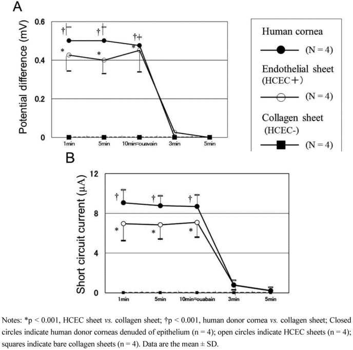
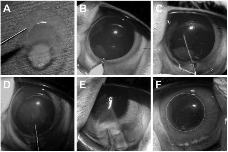
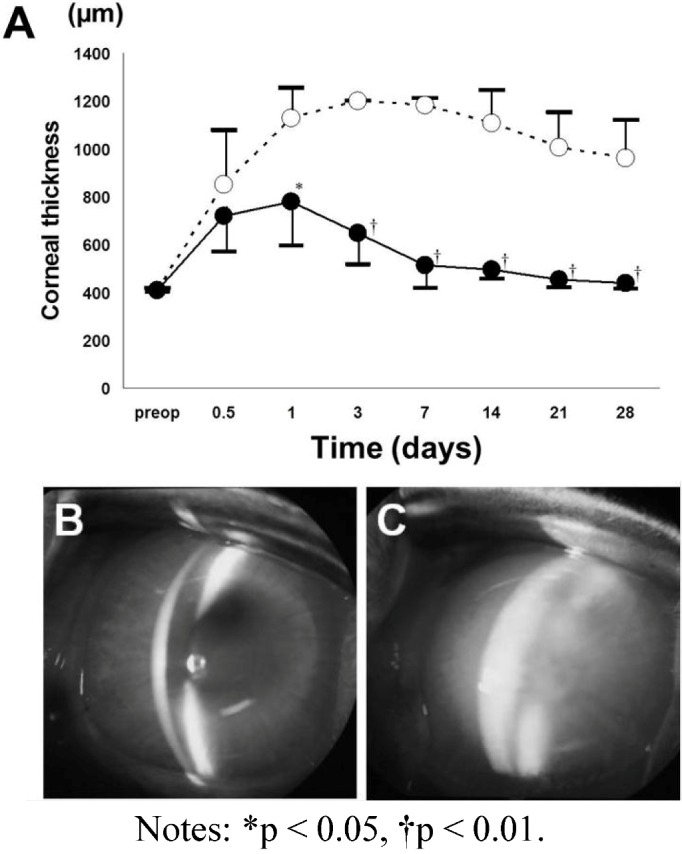
Human corneal endothelial cells (HCECs) do not replicate after wounding. Therefore, corneal endothelial deficiency can result in irreversible corneal edema. Descemet stripping automated endothelial keratoplasty (DSAEK) allows selective replacement of the diseased corneal endothelium. However, DSAEK requires a donor cornea and the worldwide shortage of corneas limits its application. This review presents current knowledge on the tissue engineering of corneal endothelium using cultured HCECs. We also provide our recent work on tissue engineering for DSAEK grafts using cultured HCECs. We reconstructed DSAEK grafts by seeding cultured DiI-labelled HCECs on collagen sheets. Then HCEC sheets were transplanted onto the posterior stroma after descemetorhexis in the DSAEK group. Severe stromal edema was detected in the control group, but not in the DSAEK group throughout the observation period. Fluorescein microscopy one month after surgery showed numerous DiI-labelled cells on the posterior corneal surface in the DSAEK group. Frozen sections showed a monolayer of DiI-labelled cells on Descemet's membrane. These findings indicate that cultured adult HCECs, transplanted with DSAEK surgery, maintain corneal transparency after transplantation and suggest the feasibility of performing DSAEK with HCECs to treat endothelial dysfunction.

摘要

人角膜内皮细胞(HCECs)受伤后不会复制。因此,角膜内皮细胞缺乏可导致不可逆的角膜水肿。后弹力层剥除自动角膜内皮移植术(DSAEK)可选择性替换病变的角膜内皮。然而,DSAEK需要供体角膜,而全球角膜短缺限制了其应用。本综述介绍了利用培养的HCECs进行角膜内皮组织工程的现有知识。我们还提供了我们最近关于使用培养的HCECs进行DSAEK移植物组织工程的工作。我们通过将培养的DiI标记的HCECs接种在胶原片上来重建DSAEK移植物。然后在DSAEK组进行后弹力层撕除后,将HCEC片移植到后基质上。在整个观察期内,对照组检测到严重的基质水肿,而DSAEK组未检测到。术后1个月荧光素显微镜检查显示DSAEK组角膜后表面有大量DiI标记的细胞。冰冻切片显示后弹力膜上有单层DiI标记的细胞。这些发现表明,通过DSAEK手术移植的培养成年HCECs在移植后可维持角膜透明度,并提示用HCECs进行DSAEK治疗内皮功能障碍的可行性。

https://cdn.ncbi.nlm.nih.gov/pmc/blobs/71ff/4030921/291599a287aa/jfb-03-00726-g006.jpg
https://cdn.ncbi.nlm.nih.gov/pmc/blobs/71ff/4030921/e826fad1fb82/jfb-03-00726-g001.jpg
https://cdn.ncbi.nlm.nih.gov/pmc/blobs/71ff/4030921/ad3f42b56f52/jfb-03-00726-g002.jpg
https://cdn.ncbi.nlm.nih.gov/pmc/blobs/71ff/4030921/884e704f4d9f/jfb-03-00726-g003.jpg
https://cdn.ncbi.nlm.nih.gov/pmc/blobs/71ff/4030921/a042b3b293f8/jfb-03-00726-g004.jpg
https://cdn.ncbi.nlm.nih.gov/pmc/blobs/71ff/4030921/f264753afae7/jfb-03-00726-g005.jpg
https://cdn.ncbi.nlm.nih.gov/pmc/blobs/71ff/4030921/291599a287aa/jfb-03-00726-g006.jpg

相似文献

1
Tissue engineering of corneal endothelium.角膜内皮的组织工程
J Funct Biomater. 2012 Oct 17;3(4):726-44. doi: 10.3390/jfb3040726.
2
Corneal endothelial regeneration and tissue engineering.角膜内皮细胞的再生与组织工程。
Prog Retin Eye Res. 2013 Jul;35:1-17. doi: 10.1016/j.preteyeres.2013.01.003. Epub 2013 Jan 23.
3
Descemet stripping automated endothelial keratoplasty using cultured corneal endothelial cells in a rabbit model.在兔模型中使用培养的角膜内皮细胞进行后弹力层剥除自动角膜内皮移植术。
Arch Ophthalmol. 2009 Oct;127(10):1321-6. doi: 10.1001/archophthalmol.2009.253.
4
Evaluation of reconstructed human corneal endothelium sheets made with porcine Descemet's membrane in vitro and in vivo.体外和体内评估使用猪 Descemet 膜制作的人角膜内皮细胞片。
Exp Eye Res. 2020 Aug;197:108125. doi: 10.1016/j.exer.2020.108125. Epub 2020 Jul 1.
5
[Transplantation of corneal endothelial cells].[角膜内皮细胞移植]
Nippon Ganka Gakkai Zasshi. 2002 Dec;106(12):805-35; discussion 836.
6
Cultured human corneal endothelial cell transplantation with a collagen sheet in a rabbit model.在兔模型中进行带胶原片的培养人角膜内皮细胞移植
Invest Ophthalmol Vis Sci. 2004 Sep;45(9):2992-7. doi: 10.1167/iovs.03-1174.
7
Transplantation of corneas reconstructed with cultured adult human corneal endothelial cells in nude rats.用培养的成年人类角膜内皮细胞重建的角膜在裸鼠中的移植。
Exp Eye Res. 2004 Aug;79(2):231-7. doi: 10.1016/j.exer.2004.05.001.
8
Corneal donor tissue preparation for endothelial keratoplasty.用于内皮角膜移植术的角膜供体组织制备
J Vis Exp. 2012 Jun 12(64):e3847. doi: 10.3791/3847.
9
Corneal donor tissue preparation for Descemet's membrane endothelial keratoplasty.用于Descemet膜内皮角膜移植术的角膜供体组织制备
J Vis Exp. 2014 Sep 17(91):51919. doi: 10.3791/51919.
10
Treatment of corneal endothelial damage in a rabbit model with a bioengineered graft using human decellularized corneal lamina and cultured human corneal endothelium.使用人去细胞角膜板层和培养的人角膜内皮细胞构建的生物工程移植物治疗兔角膜内皮损伤模型。
PLoS One. 2019 Nov 21;14(11):e0225480. doi: 10.1371/journal.pone.0225480. eCollection 2019.

引用本文的文献

1
Advances in culture, expansion and mechanistic studies of corneal endothelial cells: a systematic review.角膜内皮细胞的文化、扩增和机制研究进展:系统评价。
J Biomed Sci. 2019 Jan 4;26(1):2. doi: 10.1186/s12929-018-0492-7.
2
Corneal endothelial cell dysfunction: etiologies and management.角膜内皮细胞功能障碍:病因与治疗
Ther Adv Ophthalmol. 2018 Dec 7;10:2515841418815802. doi: 10.1177/2515841418815802. eCollection 2018 Jan-Dec.
3
Human Corneal Endothelial Cells Expanded Are a Powerful Resource for Tissue Engineering.

本文引用的文献

1
Cultivation of human corneal endothelial cells isolated from paired donor corneas.从配对供体角膜中分离培养人角膜内皮细胞。
PLoS One. 2011;6(12):e28310. doi: 10.1371/journal.pone.0028310. Epub 2011 Dec 16.
2
Increased proliferation and replicative lifespan of isolated human corneal endothelial cells with L-ascorbic acid 2-phosphate.L-抗坏血酸 2-磷酸增加分离的人角膜内皮细胞的增殖和复制寿命。
Invest Ophthalmol Vis Sci. 2011 Nov 7;52(12):8711-7. doi: 10.1167/iovs.11-7592.
3
[Physiology of the human corneal endothelium--new insights from electrophysiological investigations].
扩增后的人角膜内皮细胞是组织工程的强大资源。
Int J Med Sci. 2017 Feb 7;14(2):128-135. doi: 10.7150/ijms.17624. eCollection 2017.
4
Engineering of Human Corneal Endothelial Grafts.人角膜内皮移植片的工程化
Curr Ophthalmol Rep. 2015 Sep;3(3):207-217. doi: 10.1007/s40135-015-0077-5. Epub 2015 Jun 27.
5
Use of Mesothelial Cells and Biological Matrices for Tissue Engineering of Simple Epithelium Surrogates.间皮细胞和生物基质在简单上皮替代物组织工程中的应用。
Front Bioeng Biotechnol. 2015 Aug 17;3:117. doi: 10.3389/fbioe.2015.00117. eCollection 2015.
6
N-isopropylacrylamide-co-glycidylmethacrylate as a thermoresponsive substrate for corneal endothelial cell sheet engineering.N-异丙基丙烯酰胺-共-甲基丙烯酸缩水甘油酯作为用于角膜内皮细胞片工程的热响应性底物。
Biomed Res Int. 2014;2014:450672. doi: 10.1155/2014/450672. Epub 2014 Jun 9.
[人类角膜内皮生理学——电生理研究的新见解]
Klin Monbl Augenheilkd. 2011 Jun;228(6):520-4. doi: 10.1055/s-0031-1273254. Epub 2011 May 3.
4
A novel gelatin hydrogel carrier sheet for corneal endothelial transplantation.一种新型明胶水凝胶载体片用于角膜内皮移植。
Tissue Eng Part A. 2011 Sep;17(17-18):2213-9. doi: 10.1089/ten.TEA.2010.0568. Epub 2011 Jul 11.
5
Fabrication and characters of a corneal endothelial cells scaffold based on chitosan.基于壳聚糖的角膜内皮细胞支架的制备及特性。
J Mater Sci Mater Med. 2011 Jan;22(1):175-83. doi: 10.1007/s10856-010-4190-6. Epub 2010 Nov 25.
6
Serum-free corneal organ culture medium (SFM) but not conventional minimal essential organ culture medium (MEM) protects human corneal endothelial cells from apoptotic and necrotic cell death.无血清角膜器官培养液(SFM)而非传统的最小必需器官培养液(MEM)可保护人角膜内皮细胞免受凋亡和坏死性细胞死亡。
Br J Ophthalmol. 2011 Jan;95(1):123-30. doi: 10.1136/bjo.2010.183418. Epub 2010 Oct 24.
7
Adhesion, migration, and proliferation of cultured human corneal endothelial cells by laminin-5.层粘连蛋白-5 对培养的人眼角膜内皮细胞的黏附、迁移和增殖的影响。
Invest Ophthalmol Vis Sci. 2011 Feb 3;52(2):679-84. doi: 10.1167/iovs.10-5555.
8
Bioengineering endothelialized neo-corneas using donor-derived corneal endothelial cells and decellularized corneal stroma.利用供体来源的角膜内皮细胞和去细胞化的角膜基质生物工程化内皮neo 角膜。
Biomaterials. 2010 Sep;31(26):6738-45. doi: 10.1016/j.biomaterials.2010.05.020. Epub 2010 Jun 11.
9
TRPV channels mediate temperature-sensing in human corneal endothelial cells.瞬时受体电位香草酸通道在人眼角膜内皮细胞的温度感知中起介导作用。
Exp Eye Res. 2010 Jun;90(6):758-70. doi: 10.1016/j.exer.2010.03.010. Epub 2010 Mar 23.
10
Descemet stripping automated endothelial keratoplasty using cultured corneal endothelial cells in a rabbit model.在兔模型中使用培养的角膜内皮细胞进行后弹力层剥除自动角膜内皮移植术。
Arch Ophthalmol. 2009 Oct;127(10):1321-6. doi: 10.1001/archophthalmol.2009.253.