Feng Ju, Isern Nancy G, Burton Sarah D, Hu Jian Zhi
Pacific Northwest National Laboratory, 902 Battelle Blvd, Richland, WA 99352, USA.
Metabolites. 2013 Oct 31;3(4):1011-35. doi: 10.3390/metabo3041011.
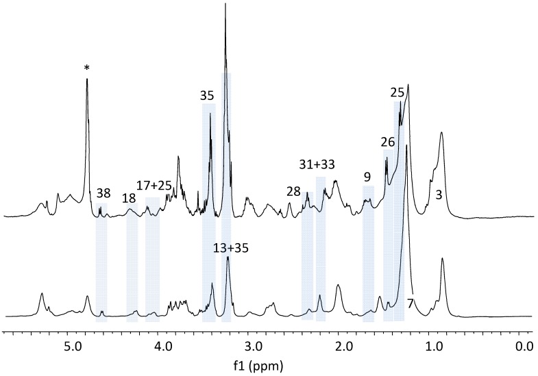
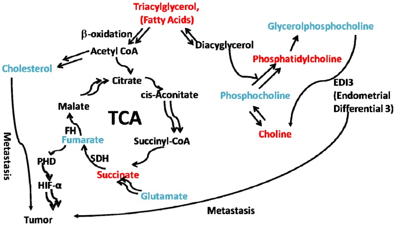
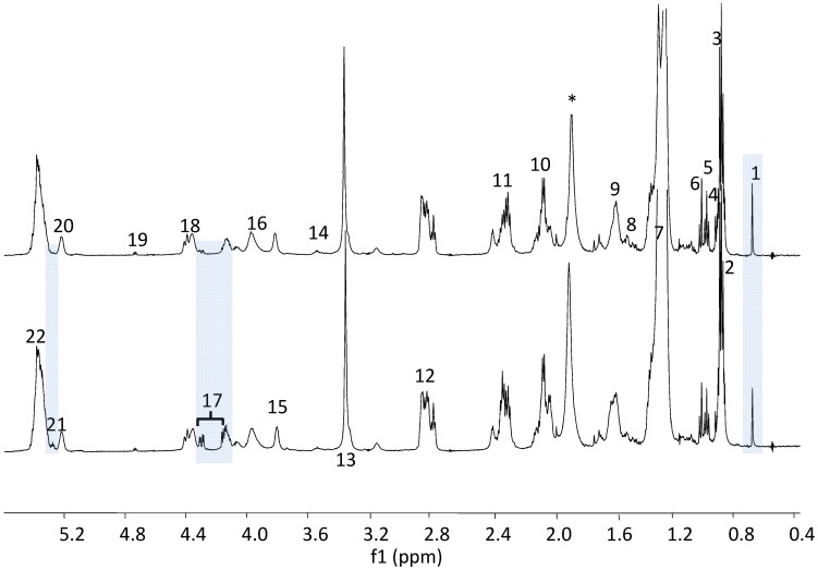
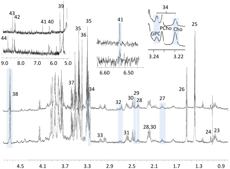
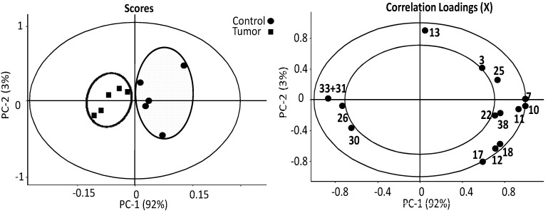
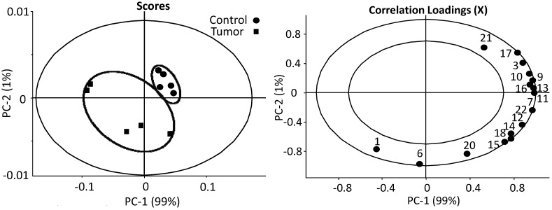
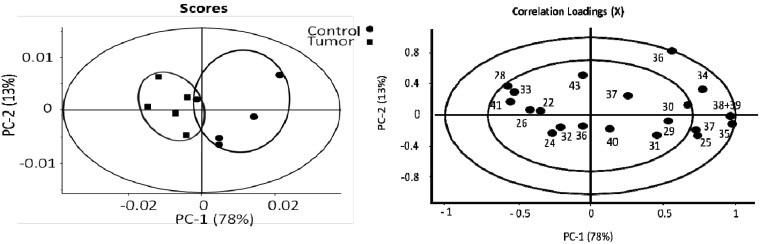
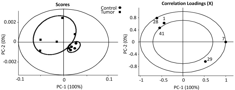
NMR metabolomics, consisting of solid state high resolution magic angle spinning (HR-MAS) 1H-NMR, liquid state high resolution 1H-NMR, and principal components analysis (PCA) has been used to study secondary metastatic B16-F10 melanoma in C57BL/6J mouse liver. The melanoma group can be differentiated from its control group by PCA analysis of the estimates of absolute concentrations from liquid state 1H-NMR spectra on liver tissue extracts or by the estimates of absolute peak intensities of metabolites from 1H HR-MAS-NMR data on intact liver tissues. In particular, we found that the estimates of absolute concentrations of glutamate, creatine, fumarate and cholesterol are elevated in the melanoma group as compared to controls, while the estimates of absolute concentrations of succinate, glycine, glucose, and the family of linear lipids including long chain fatty acids, total choline and acyl glycerol are decreased. The ratio of glycerophosphocholine (GPC) to phosphocholine (PCho) is increased by about 1.5 fold in the melanoma group, while the estimate of absolute concentration of total choline is actually lower in melanoma mice. These results suggest the following picture in secondary melanoma metastasis: Linear lipid levels are decreased by beta oxidation in the melanoma group, which contributes to an increase in the synthesis of cholesterol, and also provides an energy source input for TCA cycle. These findings suggest a link between lipid oxidation, the TCA cycle and the hypoxia-inducible factors (HIF) signal pathway in tumor metastases. Thus, this study indicates that the metabolic profile derived from NMR analysis can provide a valuable bio-signature of malignancy and cell hypoxia in metastatic melanoma.
核磁共振代谢组学,包括固态高分辨率魔角旋转(HR-MAS)1H-NMR、液态高分辨率1H-NMR和主成分分析(PCA),已被用于研究C57BL/6J小鼠肝脏中的继发性转移性B16-F10黑色素瘤。通过对肝脏组织提取物的液态1H-NMR光谱中绝对浓度估计值进行PCA分析,或通过对完整肝脏组织的1H HR-MAS-NMR数据中代谢物绝对峰强度的估计值,黑色素瘤组可与对照组区分开来。特别是,我们发现与对照组相比,黑色素瘤组中谷氨酸、肌酸、富马酸和胆固醇的绝对浓度估计值升高,而琥珀酸、甘氨酸、葡萄糖以及包括长链脂肪酸、总胆碱和酰基甘油在内的线性脂质家族的绝对浓度估计值降低。黑色素瘤组中甘油磷酸胆碱(GPC)与磷酸胆碱(PCho)的比值增加了约1.5倍,而黑色素瘤小鼠中总胆碱的绝对浓度估计值实际上更低。这些结果表明继发性黑色素瘤转移呈现出以下情况:黑色素瘤组中线性脂质水平因β氧化而降低,这有助于胆固醇合成增加,也为三羧酸循环提供能量来源输入。这些发现表明肿瘤转移中脂质氧化、三羧酸循环与缺氧诱导因子(HIF)信号通路之间存在联系。因此,本研究表明,核磁共振分析得出的代谢谱可为转移性黑色素瘤中的恶性肿瘤和细胞缺氧提供有价值的生物标志物。