Rizvi S, Yousuf S, Maheshwari V, Khan R
Institute of Ophthalmology, Jawaharlal Nehru Medical College, India.
J Surg Case Rep. 2012 Aug 1;2012(8):8. doi: 10.1093/jscr/2012.8.8.
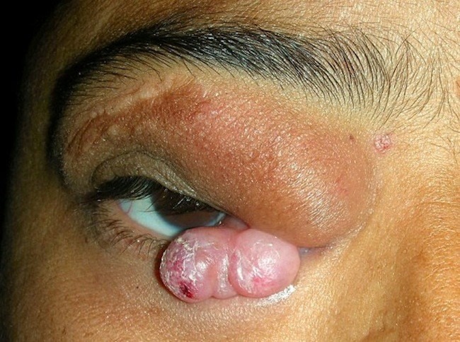
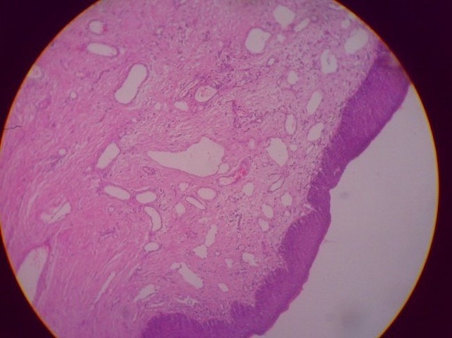
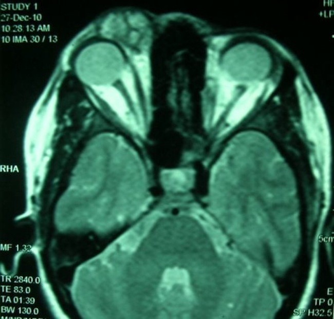
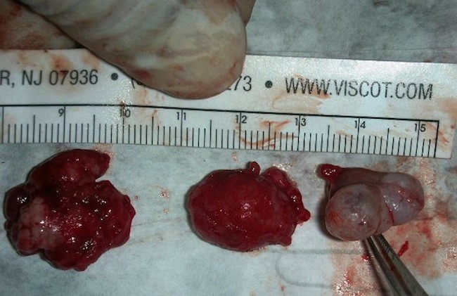
Cavernous haemangioma(CH) is mostly intraconal, single and unilateral in location. A 32 year old female presented with painless progressive growth near the inner canthus of right eye along with swelling of right upper eyelid and superomedial quadrant of the right orbit for five years. MRI showed lesions involving the preseptal space and extraconal compartment of the orbit at the superomedial aspect continous with the conjunctival swelling. Excision biopsy of the growths via anterior orbitotomy (vertical eyelid split technique) was done. Histopathological findings confirmed orbital cavernous haemangioma along with conjunctival CH. We report a rare case of multiple cavernous haemangiomas arising from the conjunctiva as well as the superomedial orbit. Complete removal of the tumour is possible even in such difficult cases as the CH is totally encapsulated and meticulous surgical dissection can give good cosmetic result to the patient.
海绵状血管瘤(CH)大多位于肌锥内,为单发且单侧性。一名32岁女性,右眼内眦附近出现无痛性进行性肿物,并伴有右上睑及右眼眶内上象限肿胀5年。磁共振成像(MRI)显示病变累及眶隔前间隙及眼眶肌锥外间隙,位于内上方,与结膜肿胀相连。通过前眶切开术(垂直睑裂技术)对肿物进行切除活检。组织病理学检查结果证实为眼眶海绵状血管瘤合并结膜CH。我们报告1例罕见的同时起源于结膜及眶内上象限的多发海绵状血管瘤病例。即使在这种困难病例中,由于CH完全被包膜包裹,完整切除肿瘤是可行的,细致的手术解剖可为患者带来良好的美容效果。