Watatani Hiroyuki, Maeshima Yohei, Hinamoto Norikazu, Yamasaki Hiroko, Ujike Haruyo, Tanabe Katsuyuki, Sugiyama Hitoshi, Otsuka Fumio, Sato Yasufumi, Makino Hirofumi
Department of Medicine and Clinical Science, Okayama University Graduate School of Medicine, Dentistry and Pharmaceutical Sciences, Okayama, 700-8558, Japan.
Department of Medicine and Clinical Science, Okayama University Graduate School of Medicine, Dentistry and Pharmaceutical Sciences, Okayama, 700-8558, Japan Department of Chronic Kidney Disease and Cardiovascular Disease, Okayama University Graduate School of Medicine, Dentistry and Pharmaceutical Sciences, Okayama, 700-8558, Japan.
Physiol Rep. 2014 Jun 27;2(6). doi: 10.14814/phy2.12054. Print 2014 Jun 1.
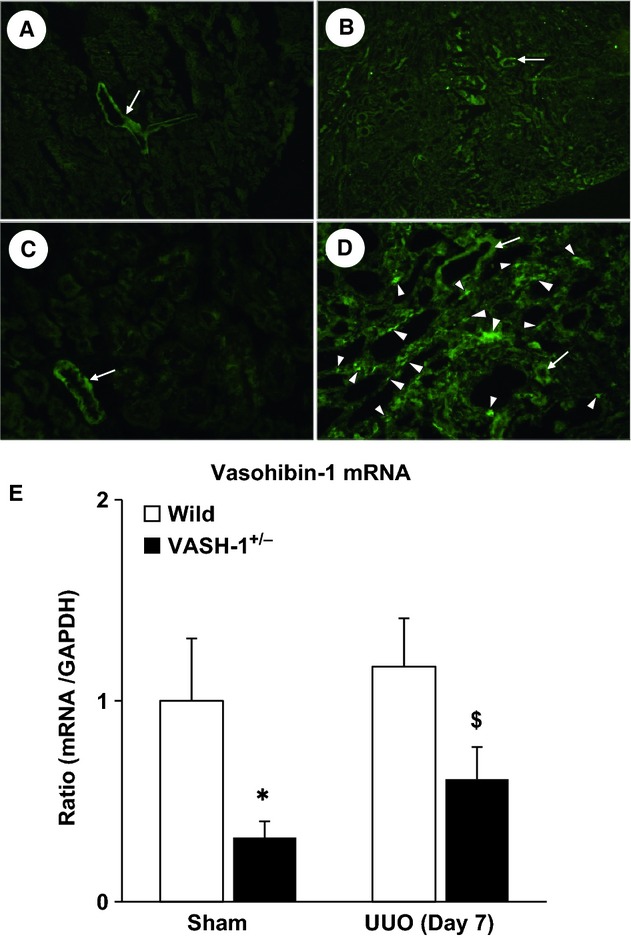
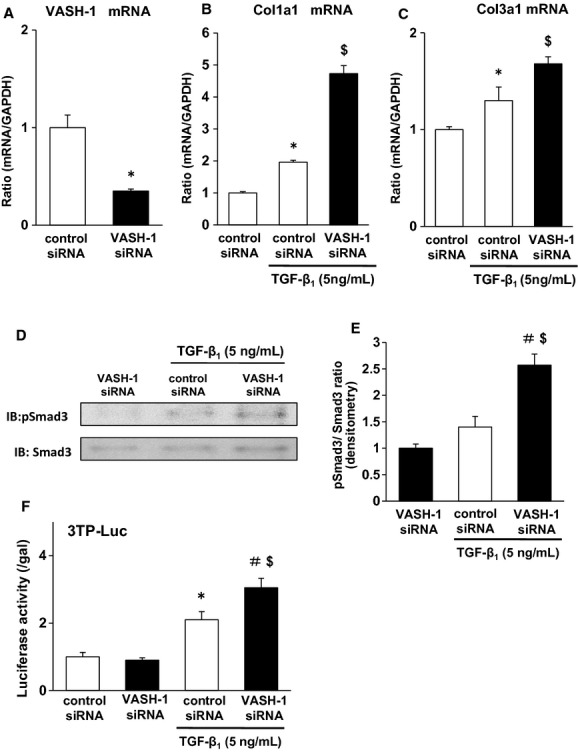
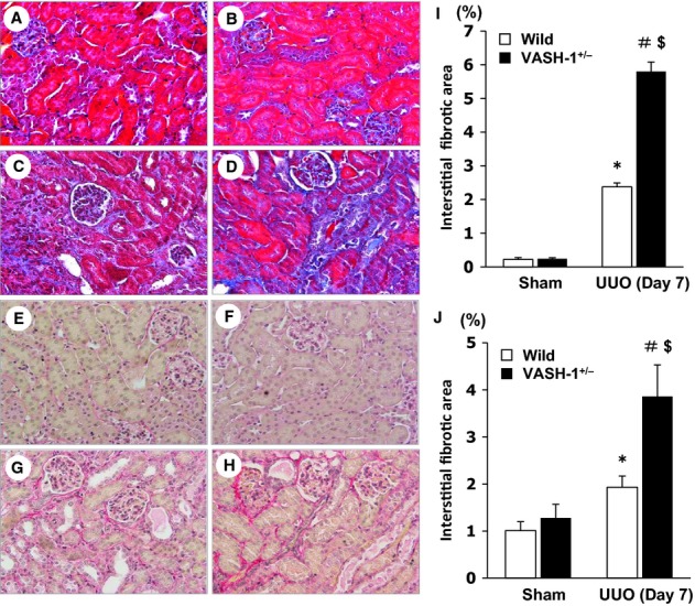
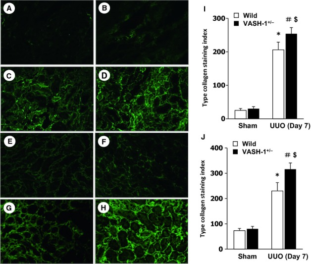
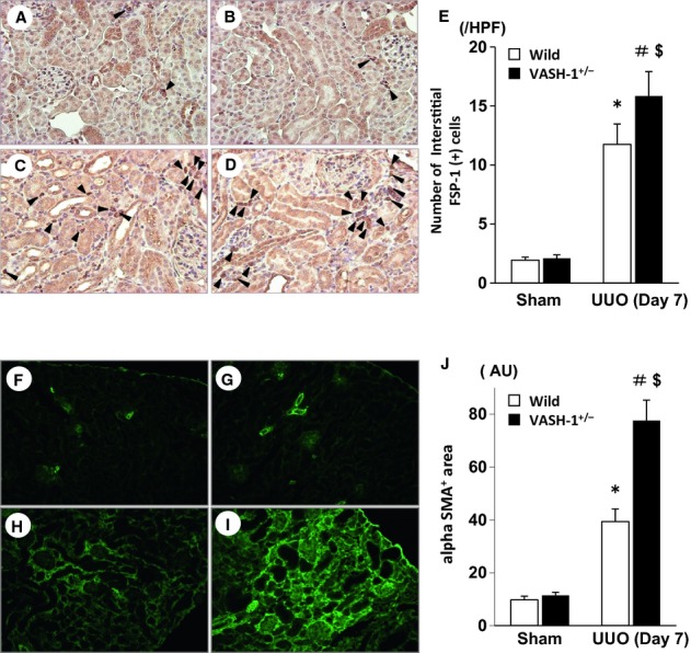
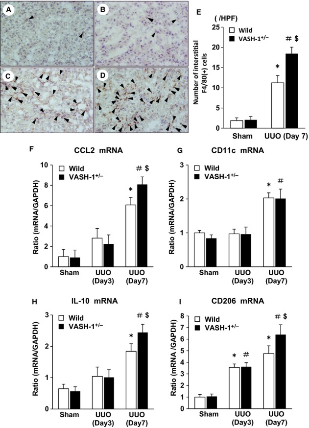
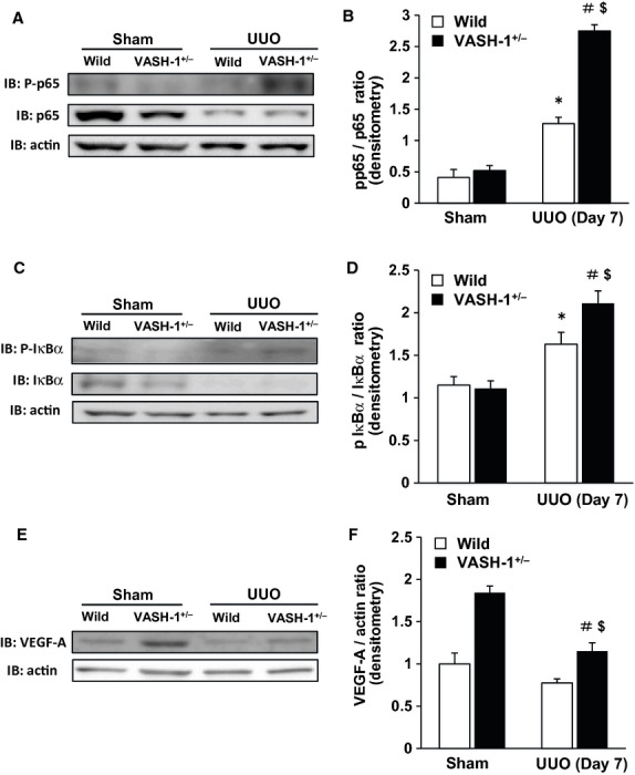
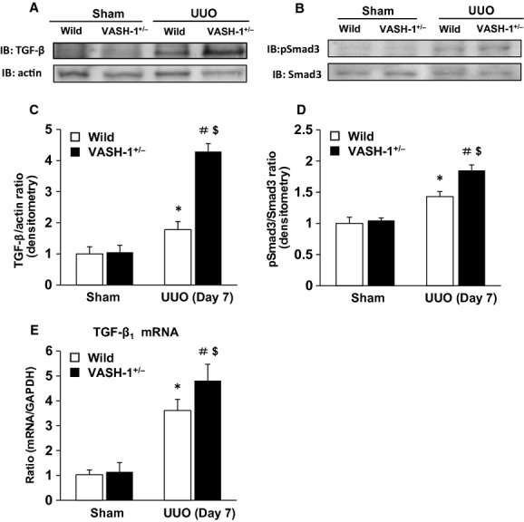
Tubulointerstitial injuries are known to predict the deterioration of renal function in chronic kidney disease (CKD). We recently reported the protective role of Vasohibin-1(VASH-1), a negative feedback regulator of angiogenesis, in diabetic nephropathy, but its impact on tubulointerstitial injuries remains to be elucidated. In the present study, we evaluated the role of endogenous VASH-1 in regulating the tubulointerstitial alterations induced by unilateral ureteral obstruction (UUO), and assessed its role on fibrogenesis and the activation of Smad3 signaling in renal fibroblasts. UUO was induced in female Vasohibin-1 heterozygous knockout mice (VASH-1(+/-)) or wild-type (WT) (VASH-1(+/+)) littermates. Mice were sacrificed on Day 7 after left ureter ligation, and the kidney tissue was obtained. Interstitial fibrosis, the accumulation of type I and type III collagen and monocytes/macrophages infiltration in the obstructed kidneys (OBK) were significantly exacerbated in VASH-1(+/-) mice compared with WT mice (Day 7). The increases in the renal levels of TGF-β1, pSmad3, NF-κB pp65, CCL2 mRNA, and the number of interstitial fibroblast-specific protein-1 (FSP-1)(+) fibroblasts in the OBK were significantly aggravated in VASH-1(+/-) mice. In addition, treatment with VASH-1 siRNA enhanced the TGF-β1-induced phosphorylation of Smad3, the transcriptional activation of the Smad3 pathway and the production of type I/type III collagen in fibroblasts, in vitro. Taken together, our findings demonstrate a protective role for endogenous VASH-1 on tubulointerstitial alterations via its regulation of inflammation and fibrosis and also show the direct anti-fibrotic effects of VASH-1 on renal fibroblasts through its modulation of TGF-β1 signaling.
已知肾小管间质损伤可预测慢性肾脏病(CKD)患者肾功能的恶化。我们最近报道了血管抑制素-1(VASH-1),一种血管生成的负反馈调节因子,在糖尿病肾病中的保护作用,但其对肾小管间质损伤的影响仍有待阐明。在本研究中,我们评估了内源性VASH-1在调节单侧输尿管梗阻(UUO)诱导的肾小管间质改变中的作用,并评估了其在肾成纤维细胞纤维化和Smad3信号激活中的作用。在雌性血管抑制素-1杂合敲除小鼠(VASH-1(+/-))或野生型(WT)(VASH-1(+/+))同窝小鼠中诱导UUO。在左侧输尿管结扎后第7天处死小鼠,获取肾脏组织。与WT小鼠相比,VASH-1(+/-)小鼠梗阻肾脏(OBK)中的间质纤维化、I型和III型胶原蛋白积累以及单核细胞/巨噬细胞浸润明显加重(第7天)。VASH-1(+/-)小鼠OBK中TGF-β1、pSmad3、NF-κB pp65、CCL2 mRNA的肾水平升高以及间质成纤维细胞特异性蛋白-1(FSP-1)(+)成纤维细胞数量明显增加。此外,在体外,用VASH-1 siRNA处理可增强TGF-β1诱导的成纤维细胞中Smad3磷酸化、Smad3途径的转录激活以及I型/III型胶原蛋白的产生。综上所述,我们的研究结果表明内源性VASH-1通过调节炎症和纤维化对肾小管间质改变具有保护作用,并且还通过调节TGF-β1信号显示VASH-1对肾成纤维细胞具有直接的抗纤维化作用。