• 文献检索
  • 文档翻译
  • 深度研究
  • 学术资讯
  • Suppr Zotero 插件Zotero 插件
  • 邀请有礼
  • 套餐&价格
  • 历史记录
应用&插件
Suppr Zotero 插件Zotero 插件浏览器插件Mac 客户端Windows 客户端微信小程序
定价
高级版会员购买积分包购买API积分包
服务
文献检索文档翻译深度研究API 文档MCP 服务
关于我们
关于 Suppr公司介绍联系我们用户协议隐私条款
关注我们

Suppr 超能文献

核心技术专利:CN118964589B侵权必究
粤ICP备2023148730 号-1Suppr @ 2026

文献检索

告别复杂PubMed语法,用中文像聊天一样搜索,搜遍4000万医学文献。AI智能推荐,让科研检索更轻松。

立即免费搜索

文件翻译

保留排版,准确专业,支持PDF/Word/PPT等文件格式,支持 12+语言互译。

免费翻译文档

深度研究

AI帮你快速写综述,25分钟生成高质量综述,智能提取关键信息,辅助科研写作。

立即免费体验

利用来自相位图的脊线和边缘的种子进行视网膜血管分割的水平集方法

Level Sets for Retinal Vasculature Segmentation Using Seeds from Ridges and Edges from Phase Maps.

作者信息

Dizdaroğlu Bekir, Ataer-Cansizoglu Esra, Kalpathy-Cramer Jayashree, Keck Katie, Chiang Michael F, Erdogmus Deniz

机构信息

Computer Engineering Department, Karadeniz Technical University, Turkey ; Cognitive Systems Laboratory, Northeastern University, Boston MA, USA.

Cognitive Systems Laboratory, Northeastern University, Boston MA, USA.

出版信息

IEEE Int Workshop Mach Learn Signal Process. 2012:1-6. doi: 10.1109/MLSP.2012.6349730.

DOI:10.1109/MLSP.2012.6349730
PMID:24975694
原文链接:https://pmc.ncbi.nlm.nih.gov/articles/PMC4071603/
Abstract

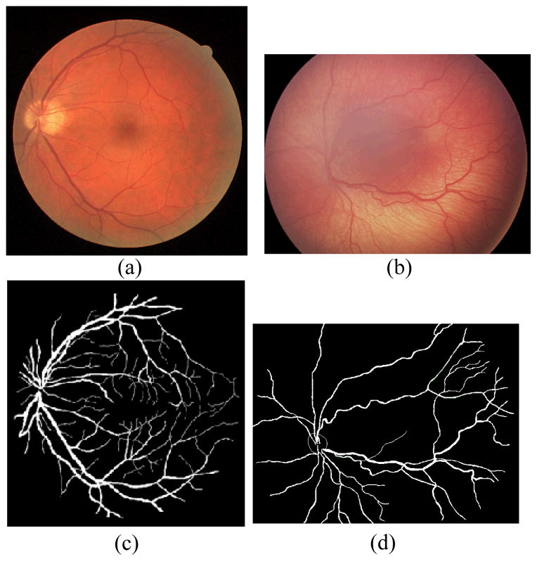
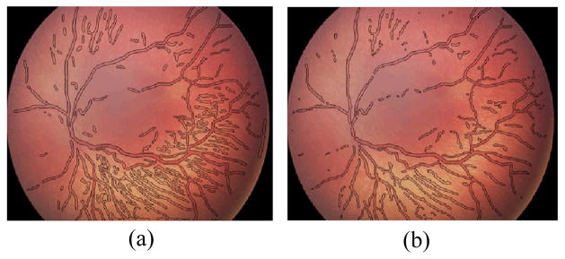
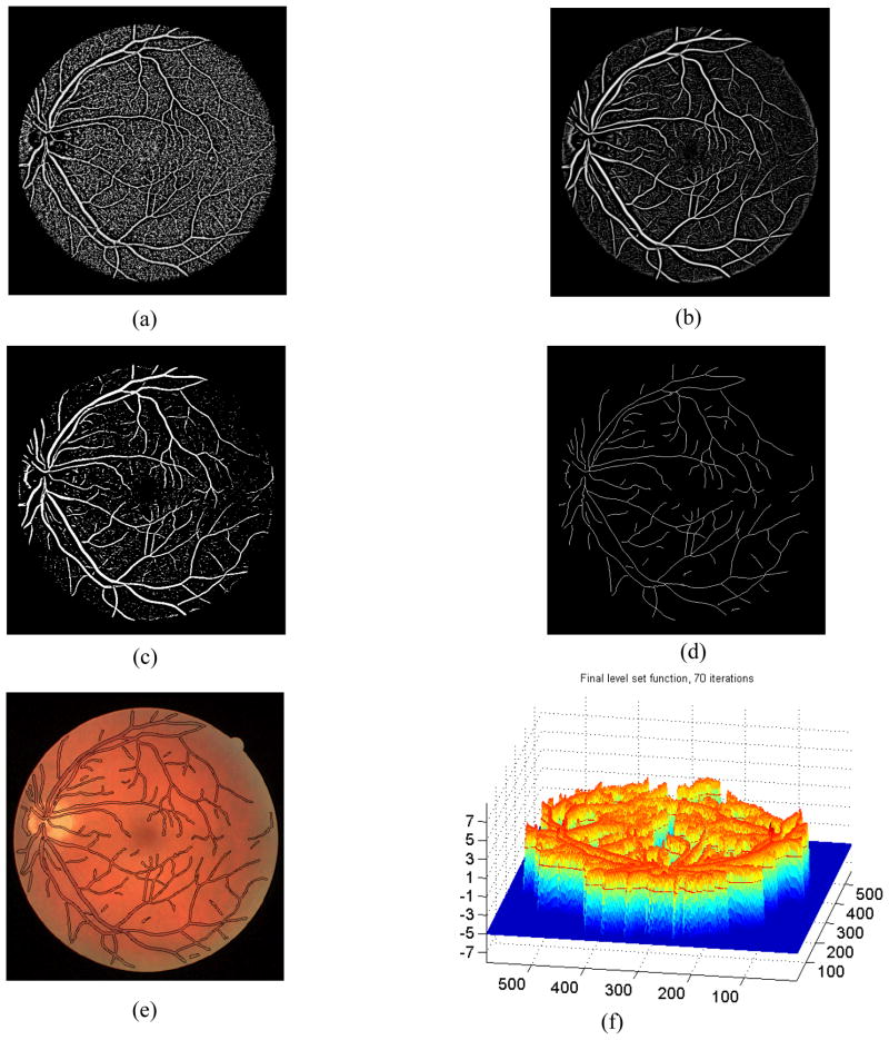
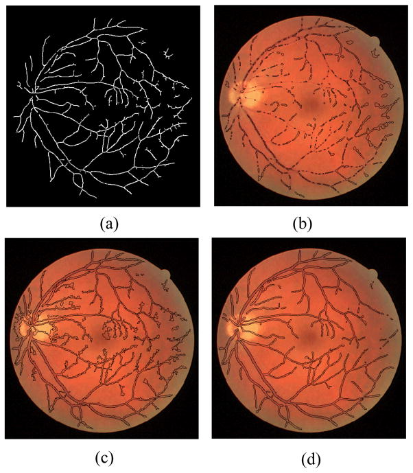
In this paper, we present a novel modification to level set based automatic retinal vasculature segmentation approaches. The method introduces ridge sample extraction for sampling the vasculature centerline and phase map based edge detection for accurate region boundary detection. Segmenting the vasculature in fundus images has been generally challenging for level set methods employing classical edge-detection methodologies. Furthermore, initialization with seed points determined by sampling vessel centerlines using ridge identification makes the method completely automated. The resulting algorithm is able to segment vasculature in fundus imagery accurately and automatically. Quantitative results supplemented with visual ones support this observation. The methodology could be applied to the broader class of vessel segmentation problems encountered in medical image analytics.

摘要

在本文中,我们提出了一种基于水平集的自动视网膜血管分割方法的新颖改进。该方法引入了用于对血管中心线进行采样的脊样本提取以及用于精确区域边界检测的基于相位图的边缘检测。对于采用经典边缘检测方法的水平集方法而言,在眼底图像中分割血管通常具有挑战性。此外,通过使用脊识别对血管中心线进行采样来确定种子点进行初始化,使得该方法完全自动化。所得算法能够准确且自动地分割眼底图像中的血管。定量结果辅以可视化结果支持了这一观察。该方法可应用于医学图像分析中遇到的更广泛的血管分割问题类别。

https://cdn.ncbi.nlm.nih.gov/pmc/blobs/1f37/4071603/7fab557fe5ed/nihms586674f4.jpg
https://cdn.ncbi.nlm.nih.gov/pmc/blobs/1f37/4071603/df4321e15cf6/nihms586674f1.jpg
https://cdn.ncbi.nlm.nih.gov/pmc/blobs/1f37/4071603/c97bfd4def26/nihms586674f2.jpg
https://cdn.ncbi.nlm.nih.gov/pmc/blobs/1f37/4071603/dbb94ee293a1/nihms586674f3.jpg
https://cdn.ncbi.nlm.nih.gov/pmc/blobs/1f37/4071603/7fab557fe5ed/nihms586674f4.jpg

相似文献

1
Level Sets for Retinal Vasculature Segmentation Using Seeds from Ridges and Edges from Phase Maps.利用来自相位图的脊线和边缘的种子进行视网膜血管分割的水平集方法
IEEE Int Workshop Mach Learn Signal Process. 2012:1-6. doi: 10.1109/MLSP.2012.6349730.
2
Interactive Blood Vessel Segmentation from Retinal Fundus Image Based on Canny Edge Detector.基于 Canny 边缘检测的视网膜眼底图像交互式血管分割。
Sensors (Basel). 2021 Sep 24;21(19):6380. doi: 10.3390/s21196380.
3
Coronary artery centerline extraction in cardiac CT angiography using a CNN-based orientation classifier.基于卷积神经网络的方向分类器的心脏 CT 血管造影中的冠状动脉中心线提取。
Med Image Anal. 2019 Jan;51:46-60. doi: 10.1016/j.media.2018.10.005. Epub 2018 Oct 22.
4
Modified pixel level snake using bottom hat transformation for evolution of retinal vasculature map.基于底部帽变换的改进像素级蛇模型在视网膜血管图演化中的应用。
Math Biosci Eng. 2021 Jun 25;18(5):5737-5757. doi: 10.3934/mbe.2021290.
5
Obtaining optic disc center and pixel region by automatic thresholding methods on morphologically processed fundus images.通过对形态学处理后的眼底图像采用自动阈值化方法获取视盘中心和像素区域。
Comput Methods Programs Biomed. 2015 Feb;118(2):173-85. doi: 10.1016/j.cmpb.2014.11.003. Epub 2014 Nov 20.
6
Automatic vasculature identification in coronary angiograms by adaptive geometrical tracking.冠状动脉造影中自适应几何跟踪的自动血管识别。
Comput Math Methods Med. 2013;2013:796342. doi: 10.1155/2013/796342. Epub 2013 Oct 22.
7
[Automatic detection of vessels in color fundus images].[彩色眼底图像中血管的自动检测]
Arch Soc Esp Oftalmol. 2010 Mar;85(3):103-9.
8
Analysis of Fundus Fluorescein Angiogram Based on the Hessian Matrix of Directional Curvelet Sub-bands and Distance Regularized Level Set Evolution.基于方向曲波子带的Hessian矩阵和距离正则化水平集演化的眼底荧光血管造影分析
J Med Signals Sens. 2015 Jul-Sep;5(3):141-55. doi: 10.4103/2228-7477.161475.
9
A new robust method for blood vessel segmentation in retinal fundus images based on weighted line detector and hidden Markov model.基于加权线检测器和隐马尔可夫模型的视网膜眼底图像血管分割新方法。
Comput Methods Programs Biomed. 2020 Apr;187:105231. doi: 10.1016/j.cmpb.2019.105231. Epub 2019 Nov 23.
10
Automated Artery Localization and Vessel Wall Segmentation using Tracklet Refinement and Polar Conversion.使用轨迹细化和极坐标转换的自动动脉定位与血管壁分割
IEEE Access. 2020;8:217603-217614. doi: 10.1109/access.2020.3040616. Epub 2020 Nov 25.

引用本文的文献

1
Retinal Vessel Extraction Assisted Multi-Channel Feature Map and U-Net.视网膜血管提取辅助多通道特征图和 U-Net
Front Public Health. 2022 Mar 17;10:858327. doi: 10.3389/fpubh.2022.858327. eCollection 2022.
2
A supervised blood vessel segmentation technique for digital Fundus images using Zernike Moment based features.基于 Zernike 矩特征的数字眼底图像有监督血管分割技术。
PLoS One. 2020 Mar 6;15(3):e0229831. doi: 10.1371/journal.pone.0229831. eCollection 2020.
3
A Multi-Anatomical Retinal Structure Segmentation System for Automatic Eye Screening Using Morphological Adaptive Fuzzy Thresholding.

本文引用的文献

1
An automated tracking approach for extraction of retinal vasculature in fundus images.一种用于眼底图像中视网膜血管提取的自动跟踪方法。
J Ophthalmic Vis Res. 2010 Jan;5(1):20-6.
2
Distance regularized level set evolution and its application to image segmentation.距离正则化水平集演化及其在图像分割中的应用。
IEEE Trans Image Process. 2010 Dec;19(12):3243-54. doi: 10.1109/TIP.2010.2069690. Epub 2010 Aug 26.
3
A new supervised method for blood vessel segmentation in retinal images by using gray-level and moment invariants-based features.
一种基于形态学自适应模糊阈值法的多解剖视网膜结构分割系统用于自动眼部筛查
IEEE J Transl Eng Health Med. 2018 May 17;6:3800123. doi: 10.1109/JTEHM.2018.2835315. eCollection 2018.
4
Robust vessel segmentation in fundus images.眼底图像中稳健的血管分割
Int J Biomed Imaging. 2013;2013:154860. doi: 10.1155/2013/154860. Epub 2013 Dec 12.
基于灰度和矩不变量特征的视网膜图像血管分割新的有监督方法。
IEEE Trans Med Imaging. 2011 Jan;30(1):146-58. doi: 10.1109/TMI.2010.2064333. Epub 2010 Aug 9.
4
Minimization of region-scalable fitting energy for image segmentation.用于图像分割的区域可缩放拟合能量最小化
IEEE Trans Image Process. 2008 Oct;17(10):1940-9. doi: 10.1109/TIP.2008.2002304.
5
Plus disease in retinopathy of prematurity: pilot study of computer-based and expert diagnosis.早产儿视网膜病变中的附加病变:基于计算机和专家诊断的初步研究
J AAPOS. 2007 Dec;11(6):532-40. doi: 10.1016/j.jaapos.2007.09.005. Epub 2007 Oct 29.
6
Interexpert agreement of plus disease diagnosis in retinopathy of prematurity.早产儿视网膜病变中加病诊断的专家间一致性
Arch Ophthalmol. 2007 Jul;125(7):875-80. doi: 10.1001/archopht.125.7.875.
7
Retinal vessel segmentation using the 2-D Gabor wavelet and supervised classification.使用二维伽柏小波和监督分类进行视网膜血管分割。
IEEE Trans Med Imaging. 2006 Sep;25(9):1214-22. doi: 10.1109/tmi.2006.879967.
8
Ridge-based vessel segmentation in color images of the retina.视网膜彩色图像中基于脊线的血管分割
IEEE Trans Med Imaging. 2004 Apr;23(4):501-9. doi: 10.1109/TMI.2004.825627.
9
Phase congruency: a low-level image invariant.
Psychol Res. 2000;64(2):136-48. doi: 10.1007/s004260000024.