Pakhomova Olga N, Gregory Betsy, Semenov Iurii, Pakhomov Andrei G
Frank Reidy Research Center for Bioelectrics, Old Dominion University, Norfolk, VA, USA.
Frank Reidy Research Center for Bioelectrics, Old Dominion University, Norfolk, VA, USA.
Biochim Biophys Acta. 2014 Oct;1838(10):2547-54. doi: 10.1016/j.bbamem.2014.06.015. Epub 2014 Jun 28.
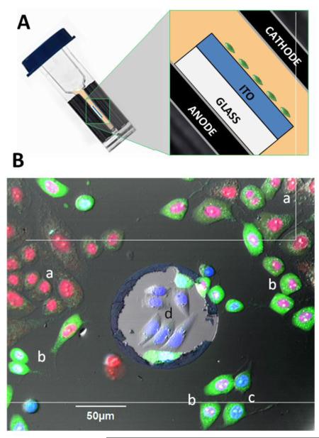
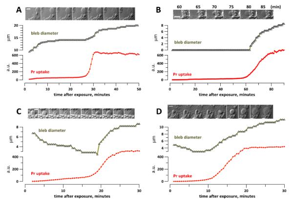
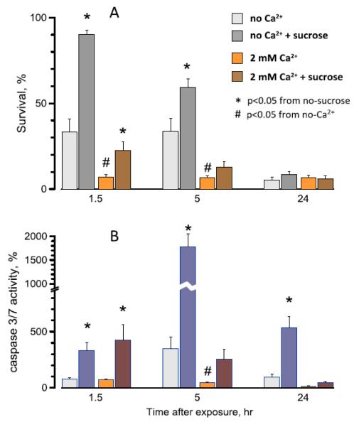
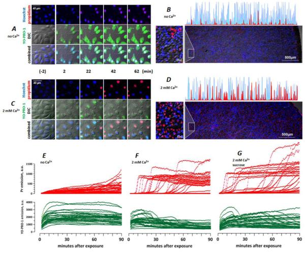
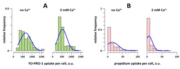
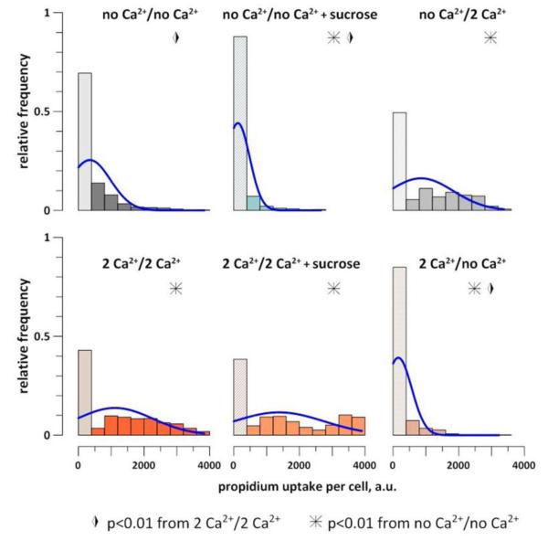
Opening of long-lived pores in the cell membrane is the principal primary effect of intense, nanosecond pulsed electric field (nsPEF). Here we demonstrate that the evolution of pores, cell survival, the time and the mode of cell death (necrotic or apoptotic) are determined by the level of external Ca(2+) after nsPEF. We also introduce a novel, minimally disruptive technique for nsEP exposure of adherent cells on indium tin oxide (ITO)-coated glass coverslips, which does not require cell detachment and enables fast exchanges of bath media. Increasing the Ca(2+) level from the nominal 2-5μM to 2mM for the first 60-90min after permeabilization by 300-nsPEF increased the early (necrotic) death in U937, CHO, and BPAE cells. With nominal Ca(2+), the inhibition of osmotic swelling rescued cells from the early necrosis and increased caspase 3/7 activation later on. However, the inhibition of swelling had a modest or no protective effect with 2mM Ca(2+) in the medium. With the nominal Ca(2+), most cells displayed gradual increase in YO-PRO-1 and propidium (Pr) uptake. With 2mM Ca(2+), the initially lower Pr uptake was eventually replaced by a massive and abrupt Pr entry (necrotic death). It was accompanied by a transient acceleration of the growth of membrane blebs due to the increase of the intracellular osmotic pressure. We conclude that the high-Ca(2+)-dependent necrotic death in nsPEF-treated cells is effected by a delayed, sudden, and osmotically-independent pore expansion (or de novo formation of larger pores), but not by the membrane rupture.
细胞膜中长寿命孔隙的开放是强纳秒脉冲电场(nsPEF)的主要初级效应。在此我们证明,nsPEF作用后,孔隙的演变、细胞存活、细胞死亡的时间和模式(坏死或凋亡)由胞外Ca(2+)水平决定。我们还介绍了一种新颖的、对铟锡氧化物(ITO)涂层玻璃盖玻片上的贴壁细胞进行nsEP暴露的微创技术,该技术无需细胞脱离,并能快速更换浴液介质。在300纳秒脉冲电场通透化处理后的最初60 - 90分钟内,将Ca(2+)水平从标称的2 - 5μM提高到2mM,会增加U937、CHO和BPAE细胞的早期(坏死)死亡。在标称Ca(2+)条件下,抑制渗透性肿胀可使细胞免于早期坏死,并在之后增加caspase 3/7的激活。然而,在培养基中Ca(2+)为2mM时,抑制肿胀的保护作用不大或没有保护作用。在标称Ca(2+)条件下,大多数细胞的YO-PRO-1和碘化丙啶(Pr)摄取逐渐增加。当Ca(2+)为2mM时,最初较低的Pr摄取最终被大量且突然的Pr进入(坏死死亡)所取代。这伴随着由于细胞内渗透压增加导致的膜泡生长的短暂加速。我们得出结论,nsPEF处理细胞中高Ca(2+)依赖性坏死死亡是由延迟的、突然的且与渗透压无关的孔隙扩张(或新形成更大的孔隙)引起的,而不是由膜破裂引起的。