Inouye Hideyo, Liu Jiliang, Makowski Lee, Palmisano Marilena, Burghammer Manfred, Riekel Christian, Kirschner Daniel A
Department of Electrical and Computer Engineering, Northeastern University, Boston, Massachusetts, United States of America.
Department of Electrical and Computer Engineering, Northeastern University, Boston, Massachusetts, United States of America; Department of Chemistry and Chemical Biology, Northeastern University, Boston, Massachusetts, United States of America.
PLoS One. 2014 Jul 1;9(7):e100592. doi: 10.1371/journal.pone.0100592. eCollection 2014.
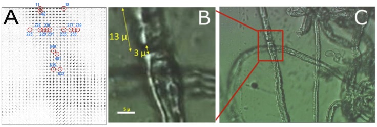
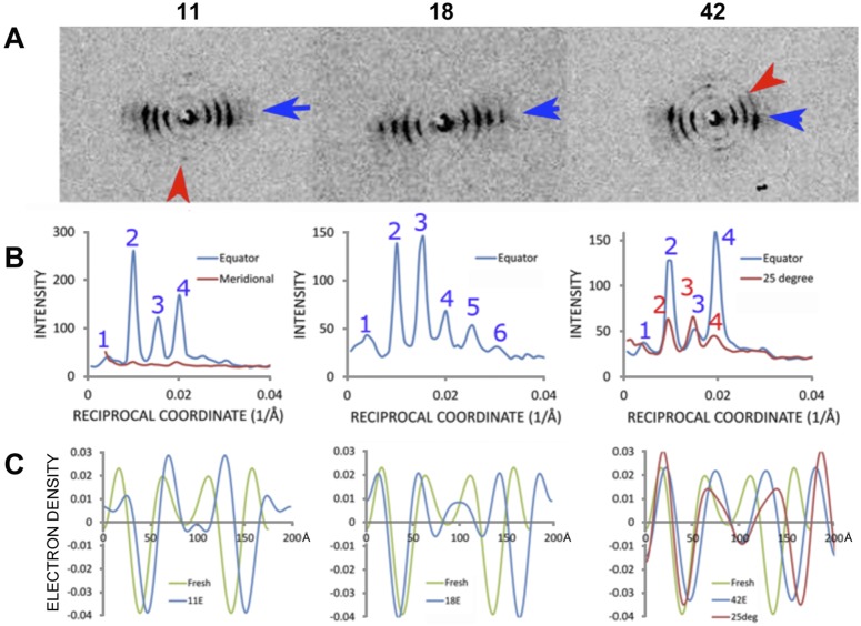
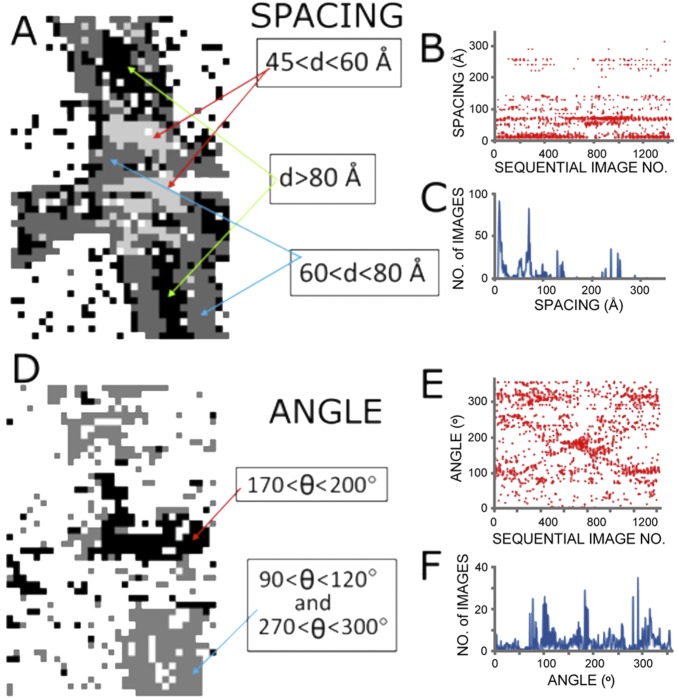
X-ray diffraction has provided extensive information about the arrangement of lipids and proteins in multilamellar myelin. This information has been limited to the abundant inter-nodal regions of the sheath because these regions dominate the scattering when x-ray beams of 100 µm diameter or more are used. Here, we used a 1 µm beam, raster-scanned across a single nerve fiber, to obtain detailed information about the molecular architecture in the nodal, paranodal, and juxtaparanodal regions. Orientation of the lamellar membrane stacks and membrane periodicity varied spatially. In the juxtaparanode-internode, 198-202 Å-period membrane arrays oriented normal to the nerve fiber axis predominated, whereas in the paranode-node, 205-208 Å-period arrays oriented along the fiber direction predominated. In parts of the sheath distal to the node, multiple sets of lamellar reflections were observed at angles to one another, suggesting that the myelin multilayers are deformed at the Schmidt-Lanterman incisures. The calculated electron density of myelin in the different regions exhibited membrane bilayer profiles with varied electron densities at the polar head groups, likely due to different amounts of major myelin proteins (P0 glycoprotein and myelin basic protein). Scattering from the center of the nerve fibers, where the x-rays are incident en face (perpendicular) to the membrane planes, provided information about the lateral distribution of protein. By underscoring the heterogeneity of membrane packing, microdiffraction analysis suggests a powerful new strategy for understanding the underlying molecular foundation of a broad spectrum of myelinopathies dependent on local specializations of myelin structure in both the PNS and CNS.
X射线衍射已提供了关于多层髓鞘中脂质和蛋白质排列的大量信息。这些信息仅限于髓鞘丰富的结间区域,因为当使用直径为100微米或更大的X射线束时,这些区域在散射中占主导地位。在这里,我们使用1微米的光束,在单根神经纤维上进行光栅扫描,以获取有关结、旁结和近旁结区域分子结构的详细信息。层状膜堆叠的方向和膜周期性在空间上有所变化。在近旁结-结间区域,垂直于神经纤维轴排列的198-202埃周期的膜阵列占主导,而在旁结-结区域,沿纤维方向排列的205-208埃周期的阵列占主导。在结远端的部分髓鞘区域,观察到多组相互成角度的层状反射,这表明髓鞘多层在施密特-兰特尔曼切迹处发生了变形。不同区域髓鞘的计算电子密度显示出膜双层轮廓,其极性头部基团处的电子密度不同,这可能是由于主要髓鞘蛋白(P0糖蛋白和髓鞘碱性蛋白)的含量不同所致。从神经纤维中心散射的X射线以正入射(垂直)于膜平面的方式提供了有关蛋白质横向分布的信息。通过强调膜堆积的异质性,微衍射分析提出了一种强有力的新策略,用于理解广泛的髓鞘病的潜在分子基础,这些髓鞘病取决于周围神经系统和中枢神经系统中髓鞘结构的局部特化。