• 文献检索
  • 文档翻译
  • 深度研究
  • 学术资讯
  • Suppr Zotero 插件Zotero 插件
  • 邀请有礼
  • 套餐&价格
  • 历史记录
应用&插件
Suppr Zotero 插件Zotero 插件浏览器插件Mac 客户端Windows 客户端微信小程序
定价
高级版会员购买积分包购买API积分包
服务
文献检索文档翻译深度研究API 文档MCP 服务
关于我们
关于 Suppr公司介绍联系我们用户协议隐私条款
关注我们

Suppr 超能文献

核心技术专利:CN118964589B侵权必究
粤ICP备2023148730 号-1Suppr @ 2026

文献检索

告别复杂PubMed语法,用中文像聊天一样搜索,搜遍4000万医学文献。AI智能推荐,让科研检索更轻松。

立即免费搜索

文件翻译

保留排版,准确专业,支持PDF/Word/PPT等文件格式,支持 12+语言互译。

免费翻译文档

深度研究

AI帮你快速写综述,25分钟生成高质量综述,智能提取关键信息,辅助科研写作。

立即免费体验

小角中子散射法获取脑切片中的神经纤维分布和髓鞘组装

Distribution and orientation of nerve fibers and myelin assembly in a brain section retrieved by small-angle neutron scattering.

机构信息

Jülich Centre of Neutron Science (JCNS-1/IBI-8), Forschungszentrum Jülich GmbH, 52425, Jülich, Germany.

Institute of Neuroscience and Medicine (INM-1), Forschungszentrum Jülich GmbH, 52425, Jülich, Germany.

出版信息

Sci Rep. 2021 Aug 27;11(1):17306. doi: 10.1038/s41598-021-92995-2.

DOI:10.1038/s41598-021-92995-2
PMID:34453063
原文链接:https://pmc.ncbi.nlm.nih.gov/articles/PMC8397781/
Abstract

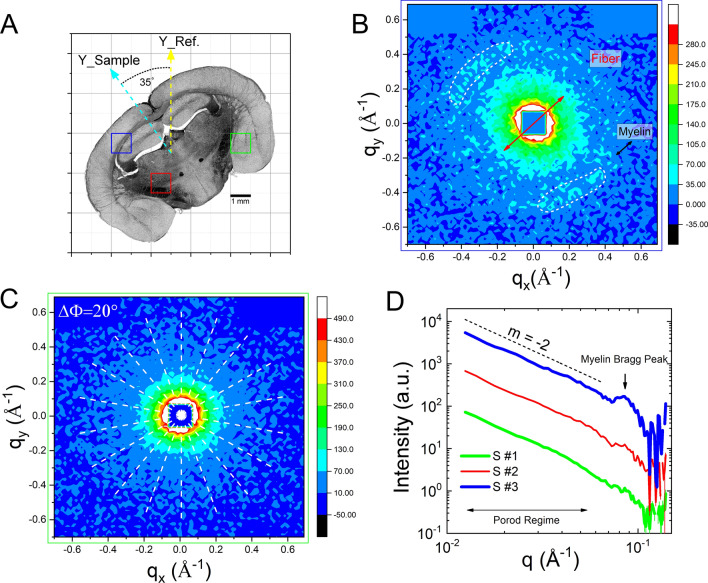
The structural connectivity of the brain has been addressed by various imaging techniques such as diffusion weighted magnetic resonance imaging (DWMRI) or specific microscopic approaches based on histological staining or label-free using polarized light (e.g., three-dimensional Polarized Light Imaging (3D-PLI), Optical Coherence Tomography (OCT)). These methods are sensitive to different properties of the fiber enwrapping myelin sheaths i.e. the distribution of myelin basic protein (histology), the apparent diffusion coefficient of water molecules restricted in their movements by the myelin sheath (DWMRI), and the birefringence of the oriented myelin lipid bilayers (3D-PLI, OCT). We show that the orientation and distribution of nerve fibers as well as myelin in thin brain sections can be determined using scanning small angle neutron scattering (sSANS). Neutrons are scattered from the fiber assembly causing anisotropic diffuse small-angle scattering and Bragg peaks related to the highly ordered periodic myelin multilayer structure. The scattering anisotropy, intensity, and angular position of the Bragg peaks can be mapped across the entire brain section. This enables mapping of the fiber and myelin distribution and their orientation in a thin brain section, which was validated by 3D-PLI. The experiments became possible by optimizing the neutron beam collimation to highest flux and enhancing the myelin contrast by deuteration. This method is very sensitive to small microstructures of biological tissue and can directly extract information on the average fiber orientation and even myelin membrane thickness. The present results pave the way toward bio-imaging for detecting structural aberrations causing neurological diseases in future.

摘要

大脑的结构连接性已经通过各种成像技术得到了研究,如扩散加权磁共振成像(DWMRI)或基于组织学染色或使用偏振光无标记的特定微观方法(例如,三维偏振光成像(3D-PLI)、光学相干断层扫描(OCT))。这些方法对包裹在髓鞘中的纤维的不同性质敏感,即髓鞘碱性蛋白的分布(组织学)、水分子在髓鞘限制下的表观扩散系数(DWMRI),以及取向的髓鞘类脂双层的双折射(3D-PLI、OCT)。我们表明,使用扫描小角中子散射(sSANS)可以确定薄脑切片中的神经纤维和髓鞘的方向和分布。中子从纤维组件散射,导致各向异性漫散射和与高度有序的周期性髓鞘多层结构相关的布拉格峰。布拉格峰的散射各向异性、强度和角度位置可以在整个脑切片上进行映射。这使得可以在薄脑切片中映射纤维和髓鞘的分布及其方向,这已通过 3D-PLI 得到验证。通过优化中子束准直以获得最高通量并通过氘化增强髓鞘对比度,使这些实验成为可能。该方法对生物组织的微小结构非常敏感,可以直接提取有关平均纤维方向甚至髓鞘膜厚度的信息。目前的结果为未来检测导致神经疾病的结构异常的生物成像铺平了道路。

https://cdn.ncbi.nlm.nih.gov/pmc/blobs/0392/8397781/3c68a6543518/41598_2021_92995_Fig7_HTML.jpg
https://cdn.ncbi.nlm.nih.gov/pmc/blobs/0392/8397781/36ffcac2c2f9/41598_2021_92995_Fig1_HTML.jpg
https://cdn.ncbi.nlm.nih.gov/pmc/blobs/0392/8397781/73a99bbe8d18/41598_2021_92995_Fig2_HTML.jpg
https://cdn.ncbi.nlm.nih.gov/pmc/blobs/0392/8397781/68021b6c253c/41598_2021_92995_Fig3_HTML.jpg
https://cdn.ncbi.nlm.nih.gov/pmc/blobs/0392/8397781/5719ea33218b/41598_2021_92995_Fig4_HTML.jpg
https://cdn.ncbi.nlm.nih.gov/pmc/blobs/0392/8397781/2d98186d8c36/41598_2021_92995_Fig5_HTML.jpg
https://cdn.ncbi.nlm.nih.gov/pmc/blobs/0392/8397781/eec2788a676d/41598_2021_92995_Fig6_HTML.jpg
https://cdn.ncbi.nlm.nih.gov/pmc/blobs/0392/8397781/3c68a6543518/41598_2021_92995_Fig7_HTML.jpg

相似文献

1
Distribution and orientation of nerve fibers and myelin assembly in a brain section retrieved by small-angle neutron scattering.小角中子散射法获取脑切片中的神经纤维分布和髓鞘组装
Sci Rep. 2021 Aug 27;11(1):17306. doi: 10.1038/s41598-021-92995-2.
2
Imaging crossing fibers in mouse, pig, monkey, and human brain using small-angle X-ray scattering.利用小角度 X 射线散射技术在小鼠、猪、猴和人脑内观察交叉纤维。
Acta Biomater. 2023 Jul 1;164:317-331. doi: 10.1016/j.actbio.2023.04.029. Epub 2023 Apr 23.
3
Retrieving neuronal orientations using 3D scanning SAXS and comparison with diffusion MRI.使用 3D 扫描 SAXS 技术获取神经元取向,并与扩散 MRI 进行比较。
Neuroimage. 2020 Jan 1;204:116214. doi: 10.1016/j.neuroimage.2019.116214. Epub 2019 Sep 27.
4
Reconstructing micrometer-scale fiber pathways in the brain: multi-contrast optical coherence tomography based tractography.重建大脑中微米级纤维通路:基于多对比度光相干断层扫描的轨迹追踪技术。
Neuroimage. 2011 Oct 15;58(4):984-92. doi: 10.1016/j.neuroimage.2011.07.005. Epub 2011 Jul 12.
5
Decoding the microstructural correlate of diffusion MRI.解析弥散磁共振成像的微观结构相关性。
NMR Biomed. 2019 Apr;32(4):e3779. doi: 10.1002/nbm.3779. Epub 2017 Aug 31.
6
Polarized light imaging of white matter architecture.白质结构的偏振光成像。
Microsc Res Tech. 2007 Oct;70(10):851-63. doi: 10.1002/jemt.20488.
7
Microstructural analysis of human white matter architecture using polarized light imaging: views from neuroanatomy.利用偏光成像技术分析人类白质结构的微观结构:神经解剖学观点。
Front Neuroinform. 2011 Nov 11;5:28. doi: 10.3389/fninf.2011.00028. eCollection 2011.
8
A novel approach to the human connectome: ultra-high resolution mapping of fiber tracts in the brain.一种新的人类连接组学方法:大脑纤维束的超高分辨率映射。
Neuroimage. 2011 Jan 15;54(2):1091-101. doi: 10.1016/j.neuroimage.2010.08.075. Epub 2010 Sep 9.
9
Structural and dynamical properties of reconstituted myelin sheaths in the presence of myelin proteins MBP and P2 studied by neutron scattering.通过中子散射研究在髓鞘蛋白MBP和P2存在下重构髓鞘的结构和动力学性质。
Soft Matter. 2014 Jan 21;10(3):519-29. doi: 10.1039/c3sm51393a.
10
A Jones matrix formalism for simulating three-dimensional polarized light imaging of brain tissue.一种用于模拟脑组织三维偏振光成像的琼斯矩阵形式。
J R Soc Interface. 2015 Oct 6;12(111):20150734. doi: 10.1098/rsif.2015.0734.

引用本文的文献

1
Interfacing with the Brain: How Nanotechnology Can Contribute.与大脑交互:纳米技术如何发挥作用。
ACS Nano. 2025 Mar 25;19(11):10630-10717. doi: 10.1021/acsnano.4c10525. Epub 2025 Mar 10.
2
Light-induced rolling of azobenzene polymer thin films for wrapping subcellular neuronal structures.用于包裹亚细胞神经元结构的偶氮苯聚合物薄膜的光诱导滚动
Commun Chem. 2024 Oct 30;7(1):249. doi: 10.1038/s42004-024-01335-8.
3
Learning from virtual experiments to assist users of Small Angle Neutron Scattering in model selection.从虚拟实验中学习以协助小角中子散射用户进行模型选择。

本文引用的文献

1
Correlative x-ray phase-contrast tomography and histology of human brain tissue affected by Alzheimer's disease.阿尔茨海默病患者人脑组织的相关 X 射线相衬断层摄影术和组织学。
Neuroimage. 2020 Apr 15;210:116523. doi: 10.1016/j.neuroimage.2020.116523. Epub 2020 Jan 11.
2
Reduced Internal Friction by Osmolyte Interaction in Intrinsically Disordered Myelin Basic Protein.通过渗透压溶质与内在无序髓鞘碱性蛋白的相互作用降低内摩擦
J Phys Chem Lett. 2020 Jan 2;11(1):292-296. doi: 10.1021/acs.jpclett.9b03001. Epub 2019 Dec 20.
3
Retrieving neuronal orientations using 3D scanning SAXS and comparison with diffusion MRI.
Sci Rep. 2024 Jul 1;14(1):14996. doi: 10.1038/s41598-024-65712-y.
4
Imaging crossing fibers in mouse, pig, monkey, and human brain using small-angle X-ray scattering.利用小角度 X 射线散射技术在小鼠、猪、猴和人脑内观察交叉纤维。
Acta Biomater. 2023 Jul 1;164:317-331. doi: 10.1016/j.actbio.2023.04.029. Epub 2023 Apr 23.
5
A multiscale tissue assessment in a rat model of mild traumatic brain injury.大鼠轻度创伤性脑损伤模型的多尺度组织评估。
J Neuropathol Exp Neurol. 2022 Dec 19;82(1):71-83. doi: 10.1093/jnen/nlac100.
使用 3D 扫描 SAXS 技术获取神经元取向,并与扩散 MRI 进行比较。
Neuroimage. 2020 Jan 1;204:116214. doi: 10.1016/j.neuroimage.2019.116214. Epub 2019 Sep 27.
4
Anomalous water dynamics in brain: a combined diffusion magnetic resonance imaging and neutron scattering investigation.脑内异常水动力学:扩散磁共振成像与中子散射的联合研究。
J R Soc Interface. 2019 Aug 30;16(157):20190186. doi: 10.1098/rsif.2019.0186. Epub 2019 Aug 14.
5
High-speed tensor tomography: iterative reconstruction tensor tomography (IRTT) algorithm.高速张量断层扫描:迭代重建张量断层扫描(IRTT)算法
Acta Crystallogr A Found Adv. 2019 Mar 1;75(Pt 2):223-238. doi: 10.1107/S2053273318017394. Epub 2019 Feb 6.
6
Voxel-wise comparisons of cellular microstructure and diffusion-MRI in mouse hippocampus using 3D Bridging of Optically-clear histology with Neuroimaging Data (3D-BOND).利用 3D-BOND(基于光透明组织学和神经影像学数据的三维桥接技术)对小鼠海马体的细胞微结构和扩散 MRI 进行体素分析比较。
Sci Rep. 2018 Mar 5;8(1):4011. doi: 10.1038/s41598-018-22295-9.
7
Developing 3D microscopy with CLARITY on human brain tissue: Towards a tool for informing and validating MRI-based histology.利用 CLARITY 在人类脑组织上开发 3D 显微镜:开发一种用于为基于 MRI 的组织学提供信息和验证的工具。
Neuroimage. 2018 Nov 15;182:417-428. doi: 10.1016/j.neuroimage.2017.11.060. Epub 2017 Nov 28.
8
A quantitative method for microstructural analysis of myelinated axons in the injured rodent brain.一种用于分析损伤后啮齿动物脑内髓鞘轴突的微观结构的定量方法。
Sci Rep. 2017 Nov 28;7(1):16492. doi: 10.1038/s41598-017-16797-1.
9
Scanning X-ray diffraction on cardiac tissue: automatized data analysis and processing.心脏组织的扫描X射线衍射:自动化数据分析与处理
J Synchrotron Radiat. 2017 Nov 1;24(Pt 6):1163-1172. doi: 10.1107/S1600577517011936. Epub 2017 Oct 23.
10
Quantifying Mesoscale Neuroanatomy Using X-Ray Microtomography.利用 X 射线微断层扫描技术定量测量介观神经解剖结构。
eNeuro. 2017 Oct 16;4(5). doi: 10.1523/ENEURO.0195-17.2017. eCollection 2017 Sep-Oct.