• 文献检索
  • 文档翻译
  • 深度研究
  • 学术资讯
  • Suppr Zotero 插件Zotero 插件
  • 邀请有礼
  • 套餐&价格
  • 历史记录
应用&插件
Suppr Zotero 插件Zotero 插件浏览器插件Mac 客户端Windows 客户端微信小程序
定价
高级版会员购买积分包购买API积分包
服务
文献检索文档翻译深度研究API 文档MCP 服务
关于我们
关于 Suppr公司介绍联系我们用户协议隐私条款
关注我们

Suppr 超能文献

核心技术专利:CN118964589B侵权必究
粤ICP备2023148730 号-1Suppr @ 2026

文献检索

告别复杂PubMed语法,用中文像聊天一样搜索,搜遍4000万医学文献。AI智能推荐,让科研检索更轻松。

立即免费搜索

文件翻译

保留排版,准确专业,支持PDF/Word/PPT等文件格式,支持 12+语言互译。

免费翻译文档

深度研究

AI帮你快速写综述,25分钟生成高质量综述,智能提取关键信息,辅助科研写作。

立即免费体验

在果蝇卵子发生过程中用于可视化肌动蛋白动力学的常见肌动蛋白标记工具的优缺点。

The pros and cons of common actin labeling tools for visualizing actin dynamics during Drosophila oogenesis.

作者信息

Spracklen Andrew J, Fagan Tiffany N, Lovander Kaylee E, Tootle Tina L

机构信息

Anatomy and Cell Biology Department, Carver College of Medicine, University of Iowa, 51 Newton Rd, Iowa City, IA 52242, USA.

Anatomy and Cell Biology Department, Carver College of Medicine, University of Iowa, 51 Newton Rd, Iowa City, IA 52242, USA.

出版信息

Dev Biol. 2014 Sep 15;393(2):209-226. doi: 10.1016/j.ydbio.2014.06.022. Epub 2014 Jul 1.

DOI:10.1016/j.ydbio.2014.06.022
PMID:24995797
原文链接:https://pmc.ncbi.nlm.nih.gov/articles/PMC4438707/
Abstract

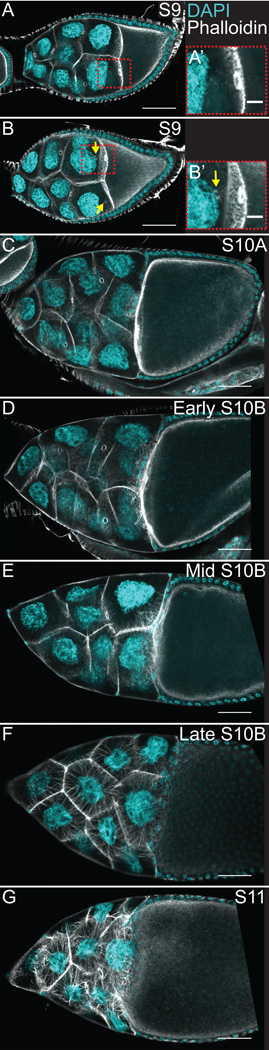
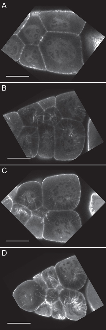
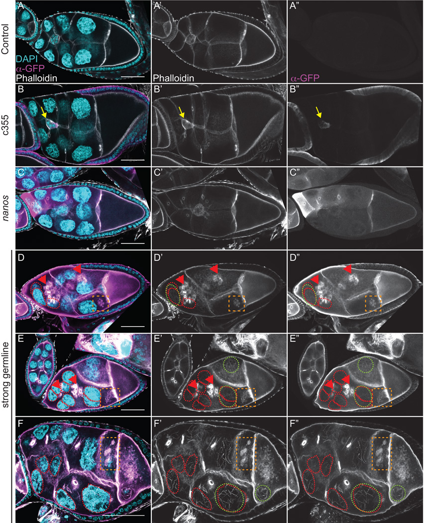
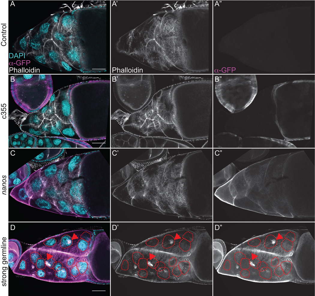
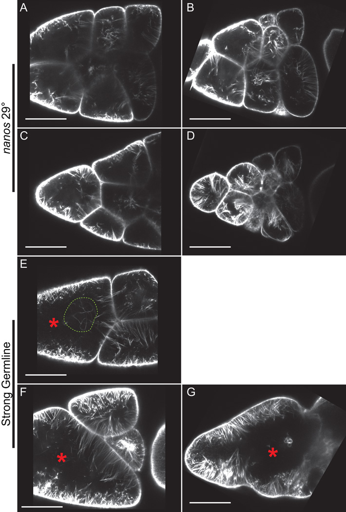
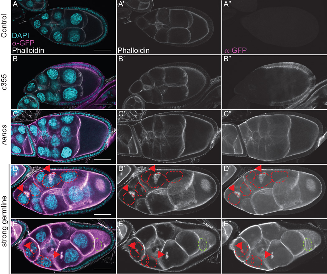
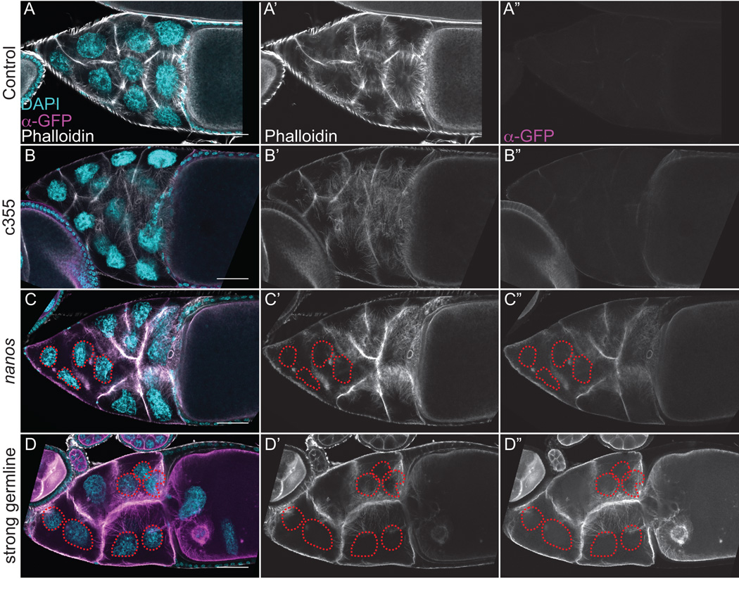
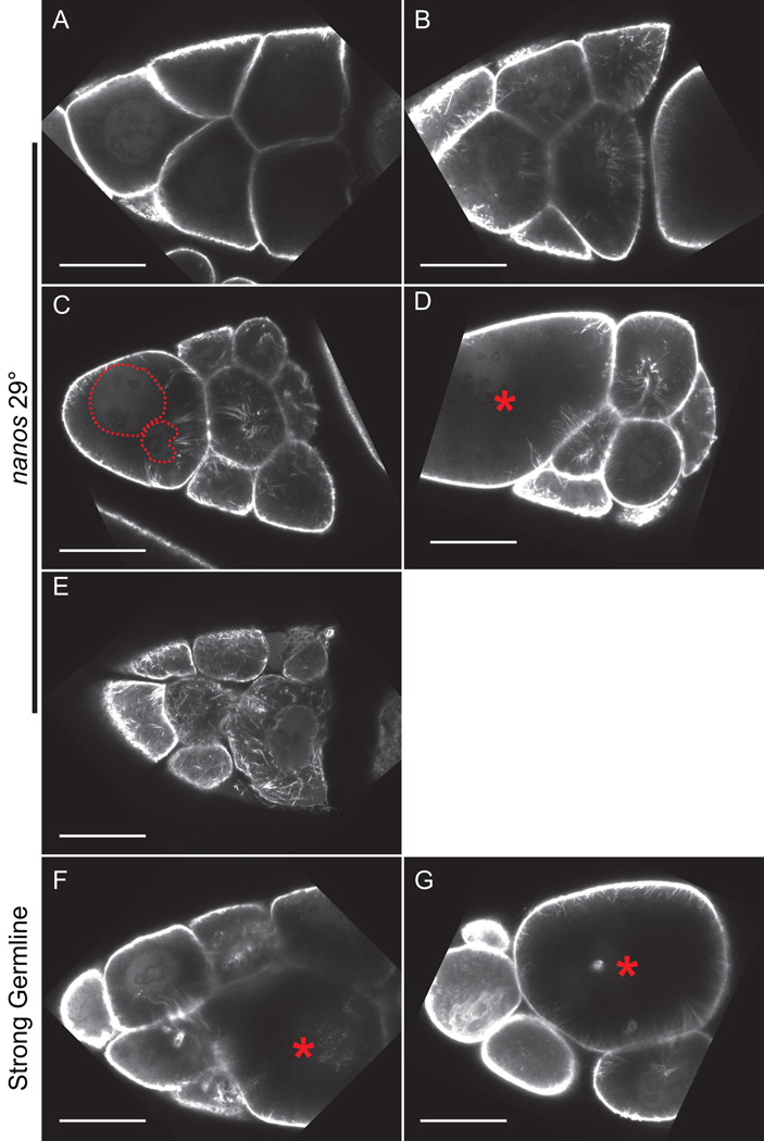
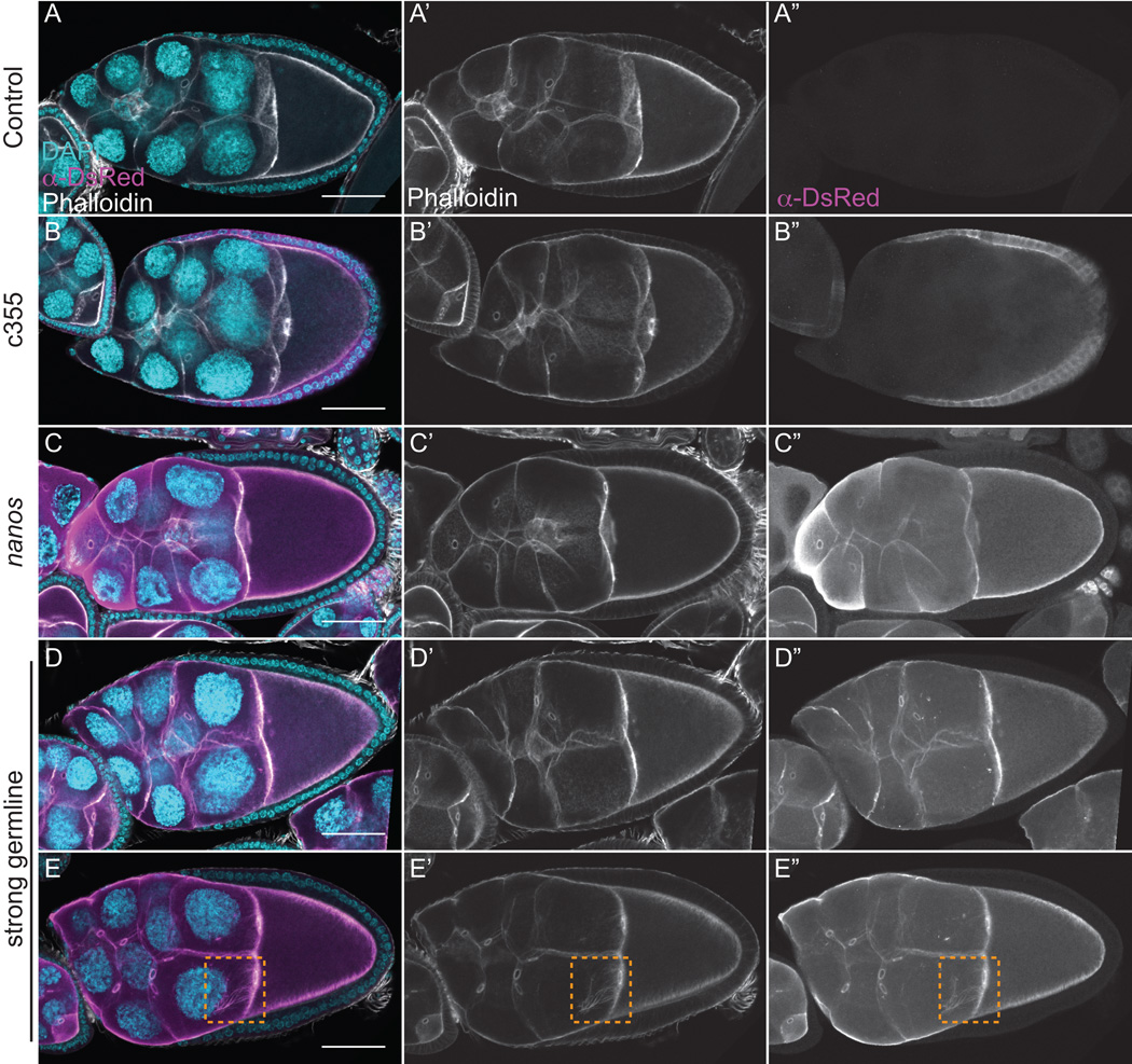
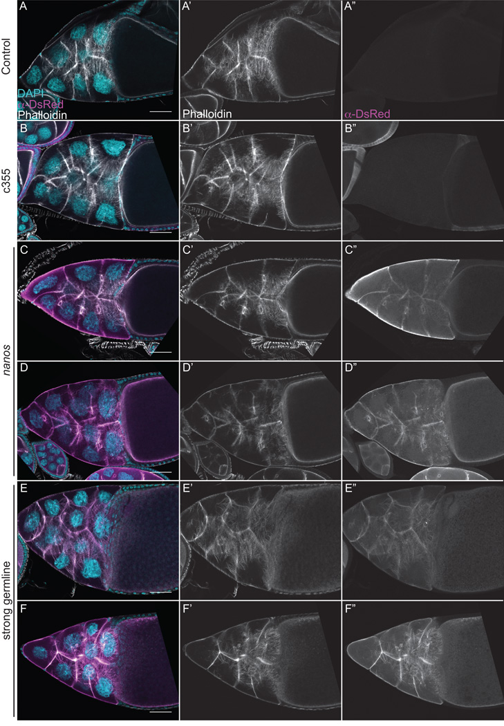
Dynamic remodeling of the actin cytoskeleton is required for both development and tissue homeostasis. While fixed image analysis has provided significant insight into such events, a complete understanding of cytoskeletal dynamics requires live imaging. Numerous tools for the live imaging of actin have been generated by fusing the actin-binding domain from an actin-interacting protein to a fluorescent protein. Here we comparatively assess the utility of three such tools--Utrophin, Lifeact, and F-tractin--for characterizing the actin remodeling events occurring within the germline-derived nurse cells during Drosophila mid-oogenesis or follicle development. Specifically, we used the UAS/GAL4 system to express these tools at different levels and in different cells, and analyzed these tools for effects on fertility, alterations in the actin cytoskeleton, and ability to label filamentous actin (F-actin) structures by both fixed and live imaging. While both Utrophin and Lifeact robustly label F-actin structures within the Drosophila germline, when strongly expressed they cause sterility and severe actin defects including cortical actin breakdown resulting in multi-nucleate nurse cells, early F-actin filament and aggregate formation during stage 9 (S9), and disorganized parallel actin filament bundles during stage 10B (S10B). However, by using a weaker germline GAL4 driver in combination with a higher temperature, Utrophin can label F-actin with minimal defects. Additionally, strong Utrophin expression within the germline causes F-actin formation in the nurse cell nuclei and germinal vesicle during mid-oogenesis. Similarly, Lifeact expression results in nuclear F-actin only within the germinal vesicle. F-tractin expresses at a lower level than the other two labeling tools, but labels cytoplasmic F-actin structures well without causing sterility or striking actin defects. Together these studies reveal how critical it is to evaluate the utility of each actin labeling tool within the tissue and cell type of interest in order to identify the tool that represents the best compromise between acceptable labeling and minimal disruption of the phenomenon being observed. In this case, we find that F-tractin, and perhaps Utrophin, when Utrophin expression levels are optimized to label efficiently without causing actin defects, can be used to study F-actin dynamics within the Drosophila nurse cells.

摘要

肌动蛋白细胞骨架的动态重塑对于发育和组织稳态都是必需的。虽然固定图像分析为这类事件提供了重要见解,但要全面理解细胞骨架动力学则需要实时成像。通过将肌动蛋白相互作用蛋白的肌动蛋白结合结构域与荧光蛋白融合,已经产生了许多用于肌动蛋白实时成像的工具。在这里,我们比较评估了三种这样的工具——肌萎缩蛋白相关蛋白(Utrophin)、生命肌动蛋白(Lifeact)和F - 肌动蛋白(F-tractin)——在果蝇卵子发生中期或卵泡发育过程中,用于表征生殖系来源的滋养细胞内发生的肌动蛋白重塑事件的效用。具体而言,我们使用UAS/GAL4系统在不同水平和不同细胞中表达这些工具,并通过固定和实时成像分析这些工具对生育力的影响、肌动蛋白细胞骨架的改变以及标记丝状肌动蛋白(F-肌动蛋白)结构的能力。虽然肌萎缩蛋白相关蛋白和生命肌动蛋白都能有力地标记果蝇生殖系中的F-肌动蛋白结构,但当它们强烈表达时会导致不育和严重的肌动蛋白缺陷,包括皮质肌动蛋白分解导致多核滋养细胞、在第9阶段(S9)早期形成F-肌动蛋白丝和聚集体,以及在第10B阶段(S10B)时平行肌动蛋白丝束紊乱。然而,通过使用较弱的生殖系GAL4驱动子并结合较高温度,肌萎缩蛋白相关蛋白可以以最小的缺陷标记F-肌动蛋白。此外,生殖系中强烈的肌萎缩蛋白相关蛋白表达会在卵子发生中期导致滋养细胞核和生发泡中形成F-肌动蛋白。同样,生命肌动蛋白的表达仅在生发泡内导致核F-肌动蛋白形成。F - 肌动蛋白的表达水平低于其他两种标记工具,但能很好地标记细胞质F-肌动蛋白结构,且不会导致不育或明显的肌动蛋白缺陷。这些研究共同揭示了在感兴趣的组织和细胞类型中评估每种肌动蛋白标记工具的效用是多么关键,以便确定在可接受的标记和对所观察现象的最小干扰之间达到最佳平衡的工具。在这种情况下,我们发现F - 肌动蛋白,也许还有肌萎缩蛋白相关蛋白(当肌萎缩蛋白相关蛋白的表达水平经过优化以有效标记而不引起肌动蛋白缺陷时),可用于研究果蝇滋养细胞内的F-肌动蛋白动力学。

https://cdn.ncbi.nlm.nih.gov/pmc/blobs/d490/4438707/63f7cbef105a/nihms689377f10.jpg
https://cdn.ncbi.nlm.nih.gov/pmc/blobs/d490/4438707/95526c405684/nihms689377f1.jpg
https://cdn.ncbi.nlm.nih.gov/pmc/blobs/d490/4438707/4eab54e405d7/nihms689377f2.jpg
https://cdn.ncbi.nlm.nih.gov/pmc/blobs/d490/4438707/bb0ab48fc798/nihms689377f3.jpg
https://cdn.ncbi.nlm.nih.gov/pmc/blobs/d490/4438707/04674760c0c3/nihms689377f4.jpg
https://cdn.ncbi.nlm.nih.gov/pmc/blobs/d490/4438707/6a0c72406d5c/nihms689377f5.jpg
https://cdn.ncbi.nlm.nih.gov/pmc/blobs/d490/4438707/a82dffa10516/nihms689377f6.jpg
https://cdn.ncbi.nlm.nih.gov/pmc/blobs/d490/4438707/139740a6ba25/nihms689377f7.jpg
https://cdn.ncbi.nlm.nih.gov/pmc/blobs/d490/4438707/7f16e890e287/nihms689377f8.jpg
https://cdn.ncbi.nlm.nih.gov/pmc/blobs/d490/4438707/e61bf89d847e/nihms689377f9.jpg
https://cdn.ncbi.nlm.nih.gov/pmc/blobs/d490/4438707/63f7cbef105a/nihms689377f10.jpg

相似文献

1
The pros and cons of common actin labeling tools for visualizing actin dynamics during Drosophila oogenesis.在果蝇卵子发生过程中用于可视化肌动蛋白动力学的常见肌动蛋白标记工具的优缺点。
Dev Biol. 2014 Sep 15;393(2):209-226. doi: 10.1016/j.ydbio.2014.06.022. Epub 2014 Jul 1.
2
Prostaglandins temporally regulate cytoplasmic actin bundle formation during Drosophila oogenesis.前列腺素在果蝇卵子发生过程中暂时调节细胞质肌动蛋白束的形成。
Mol Biol Cell. 2014 Feb;25(3):397-411. doi: 10.1091/mbc.E13-07-0366. Epub 2013 Nov 27.
3
Comparative analysis of tools for live cell imaging of actin network architecture.肌动蛋白网络结构活细胞成像工具的比较分析
Bioarchitecture. 2014;4(6):189-202. doi: 10.1080/19490992.2014.1047714. Epub 2015 Aug 28.
4
Hsp60C is required in follicle as well as germline cells during oogenesis in Drosophila melanogaster.在黑腹果蝇卵子发生过程中,Hsp60C在卵泡以及生殖细胞中是必需的。
Dev Dyn. 2008 May;237(5):1334-47. doi: 10.1002/dvdy.21524.
5
HtsRC-Mediated Accumulation of F-Actin Regulates Ring Canal Size During Oogenesis.HtsRC 介导的 F-肌动蛋白积累调控卵子发生过程中的环道大小。
Genetics. 2020 Nov;216(3):717-734. doi: 10.1534/genetics.120.303629. Epub 2020 Sep 3.
6
Affimer proteins for F-actin: novel affinity reagents that label F-actin in live and fixed cells.Affimer 蛋白与 F-actin:新型亲和试剂,可标记活细胞和固定细胞中的 F-actin。
Sci Rep. 2018 Apr 26;8(1):6572. doi: 10.1038/s41598-018-24953-4.
7
The Drosophila RNA-binding protein Lark is required for the organization of the actin cytoskeleton and Hu-li tai shao localization during oogenesis.果蝇RNA结合蛋白Lark在卵子发生过程中对于肌动蛋白细胞骨架的组织和“糊涂太少”定位是必需的。
Genesis. 2004 Oct;40(2):90-100. doi: 10.1002/gene.20069.
8
Domains of the Pavarotti kinesin-like protein that direct its subcellular distribution: effects of mislocalisation on the tubulin and actin cytoskeleton during Drosophila oogenesis.帕瓦罗蒂类驱动蛋白决定其亚细胞分布的结构域:果蝇卵子发生过程中定位错误对微管蛋白和肌动蛋白细胞骨架的影响
J Cell Sci. 2002 Feb 15;115(Pt 4):725-36. doi: 10.1242/jcs.115.4.725.
9
Pharmaco-Genetic Screen To Uncover Actin Regulators Targeted by Prostaglandins During Oogenesis.药物遗传学筛选以揭示卵子发生过程中前列腺素靶向的肌动蛋白调节因子。
G3 (Bethesda). 2019 Nov 5;9(11):3555-3565. doi: 10.1534/g3.119.400704.
10
Visualization of Actin Cytoskeletal Dynamics in Fixed and Live Drosophila Egg Chambers.固定和活的果蝇卵室中肌动蛋白细胞骨架动力学的可视化
Methods Mol Biol. 2015;1328:113-24. doi: 10.1007/978-1-4939-2851-4_8.

引用本文的文献

1
ADPact: A versatile sensor for ADP-F-actin.ADPact:一种用于ADP-丝状肌动蛋白的多功能传感器。
Proc Natl Acad Sci U S A. 2025 Jul 22;122(29):e2420467122. doi: 10.1073/pnas.2420467122. Epub 2025 Jul 16.
2
Nonmuscle myosin II regulates presynaptic actin and neuronal mechanobiology in Drosophila.非肌肉肌球蛋白II调节果蝇突触前肌动蛋白和神经元力学生物学。
J Cell Biol. 2025 Sep 1;224(9). doi: 10.1083/jcb.202501211. Epub 2025 Jul 11.
3
Prostaglandins limit nuclear actin rod formation during oogenesis.前列腺素在卵子发生过程中限制核肌动蛋白杆的形成。

本文引用的文献

1
Prostaglandins temporally regulate cytoplasmic actin bundle formation during Drosophila oogenesis.前列腺素在果蝇卵子发生过程中暂时调节细胞质肌动蛋白束的形成。
Mol Biol Cell. 2014 Feb;25(3):397-411. doi: 10.1091/mbc.E13-07-0366. Epub 2013 Nov 27.
2
Gβ1 controls collective cell migration by regulating the protrusive activity of leader cells in the posterior lateral line primordium.Gβ1 通过调节后外侧线原基中先导细胞的伸出活性来控制细胞的集体迁移。
Dev Biol. 2014 Jan 15;385(2):316-27. doi: 10.1016/j.ydbio.2013.10.027. Epub 2013 Nov 4.
3
Filopodia-like actin cables position nuclei in association with perinuclear actin in Drosophila nurse cells.
MicroPubl Biol. 2025 Apr 3;2025. doi: 10.17912/micropub.biology.001571. eCollection 2025.
4
Cell deformations generated by stochastic actomyosin waves drive in vivo random-walk swimming migration.由随机肌动球蛋白波产生的细胞变形驱动体内随机游走式游动迁移。
J Cell Sci. 2025 May 15;138(10). doi: 10.1242/jcs.263787. Epub 2025 May 22.
5
Structure of the F-tractin-F-actin complex.F-驱动蛋白-F-肌动蛋白复合物的结构。
J Cell Biol. 2025 Apr 7;224(4). doi: 10.1083/jcb.202409192. Epub 2025 Feb 10.
6
Moesin integrates cortical and lamellar actin networks during Drosophila macrophage migration.在果蝇巨噬细胞迁移过程中,膜突蛋白整合皮质肌动蛋白网络和片层肌动蛋白网络。
Nat Commun. 2025 Feb 6;16(1):1414. doi: 10.1038/s41467-024-55510-5.
7
Design and Use of AsLOV2-Based Optogenetic Tools for Actin Imaging.用于肌动蛋白成像的基于AsLOV2的光遗传学工具的设计与应用
Methods Mol Biol. 2025;2840:89-100. doi: 10.1007/978-1-0716-4047-0_7.
8
Nuclear actin is a critical regulator of female germline stem cell maintenance.核肌动蛋白是雌性生殖系干细胞维持的关键调节因子。
bioRxiv. 2024 Aug 28:2024.08.27.609996. doi: 10.1101/2024.08.27.609996.
9
Diaph1 knockout inhibits mouse primordial germ cell proliferation and affects gonadal development.Diaph1 敲除抑制小鼠原始生殖细胞增殖并影响性腺发育。
Reprod Biol Endocrinol. 2024 Jul 15;22(1):82. doi: 10.1186/s12958-024-01257-z.
10
Detection of fluorescent protein mechanical switching in cellulo.在细胞内检测荧光蛋白的机械开关。
Cell Rep Methods. 2024 Jul 15;4(7):100815. doi: 10.1016/j.crmeth.2024.100815. Epub 2024 Jul 9.
丝状伪足状肌动蛋白纤维与核周肌动蛋白一起定位细胞核,这与果蝇滋养细胞中有关。
Dev Cell. 2013 Sep 30;26(6):604-15. doi: 10.1016/j.devcel.2013.08.014.
4
To be or not to be assembled: progressing into nuclear actin filaments.存在或不存在组装:进入核肌动蛋白丝。
Nat Rev Mol Cell Biol. 2013 Nov;14(11):693-7. doi: 10.1038/nrm3681. Epub 2013 Oct 3.
5
Filopodia are dispensable for endothelial tip cell guidance.丝状伪足对于血管内皮细胞前体细胞的导向作用可有可无。
Development. 2013 Oct;140(19):4031-40. doi: 10.1242/dev.097352.
6
CIP4 coordinates with phospholipids and actin-associated proteins to localize to the protruding edge and produce actin ribs and veils.CIP4 与磷脂和肌动蛋白相关蛋白协调定位到突出的边缘,并产生肌动蛋白肋骨和面纱。
J Cell Sci. 2013 Jun 1;126(Pt 11):2411-23. doi: 10.1242/jcs.117473. Epub 2013 Apr 9.
7
Nuclear actin network assembly by formins regulates the SRF coactivator MAL.通过形成蛋白调节核肌动蛋白网络组装,从而调控血清反应因子共激活因子 MAL。
Science. 2013 May 17;340(6134):864-7. doi: 10.1126/science.1235038. Epub 2013 Apr 4.
8
Visualization of actin filaments and monomers in somatic cell nuclei.体细胞细胞核中肌动蛋白丝和单体的可视化。
Mol Biol Cell. 2013 Apr;24(7):982-94. doi: 10.1091/mbc.E12-09-0685. Epub 2013 Feb 27.
9
Drosophila Fascin is a novel downstream target of prostaglandin signaling during actin remodeling.果蝇 Fascin 是前列腺素信号在肌动蛋白重塑过程中的一个新的下游靶标。
Mol Biol Cell. 2012 Dec;23(23):4567-78. doi: 10.1091/mbc.E12-05-0417. Epub 2012 Oct 10.
10
Transcriptional regulation of Profilin during wound closure in Drosophila larvae.果蝇幼虫伤口闭合过程中 Profilin 的转录调控。
J Cell Sci. 2012 Dec 1;125(Pt 23):5667-76. doi: 10.1242/jcs.107490. Epub 2012 Sep 12.