Kelly Christopher V, Wakefield Devin L, Holowka David A, Craighead Harold G, Baird Barbara A
Department of Chemistry and Chemical Biology, Cornell University , Ithaca, New York 14853, United States.
ACS Nano. 2014 Jul 22;8(7):7392-404. doi: 10.1021/nn502593k. Epub 2014 Jul 11.
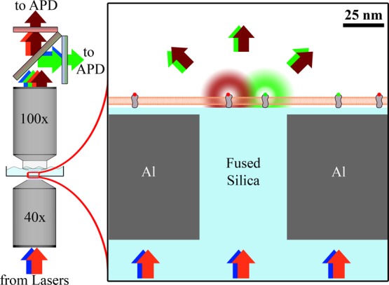
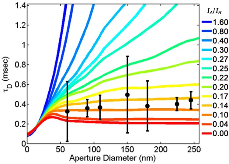
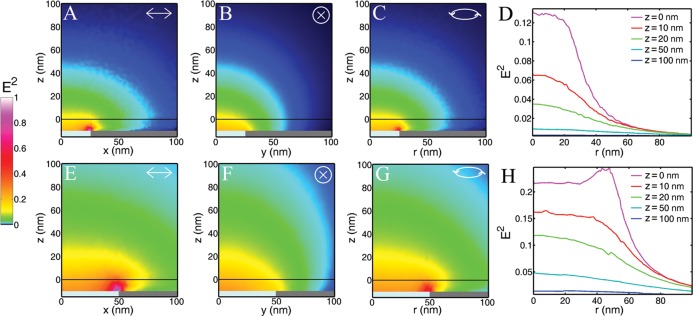
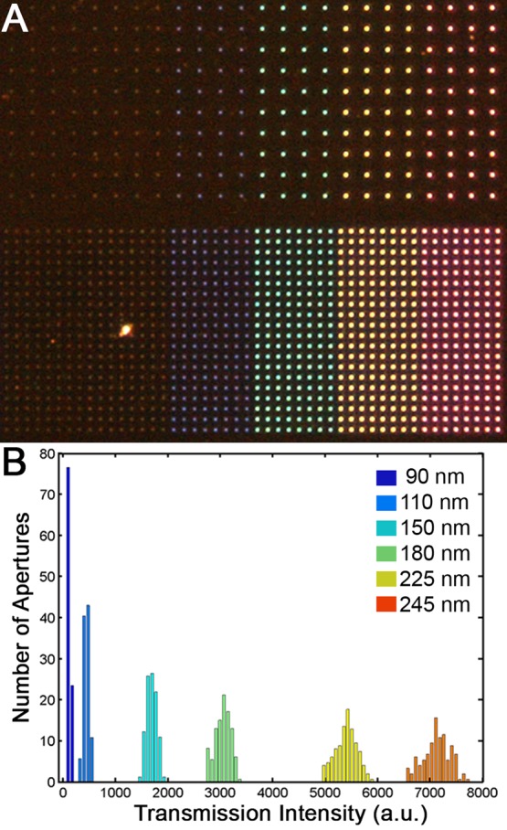
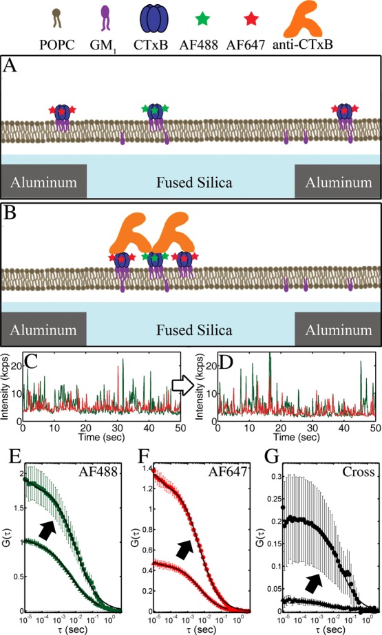
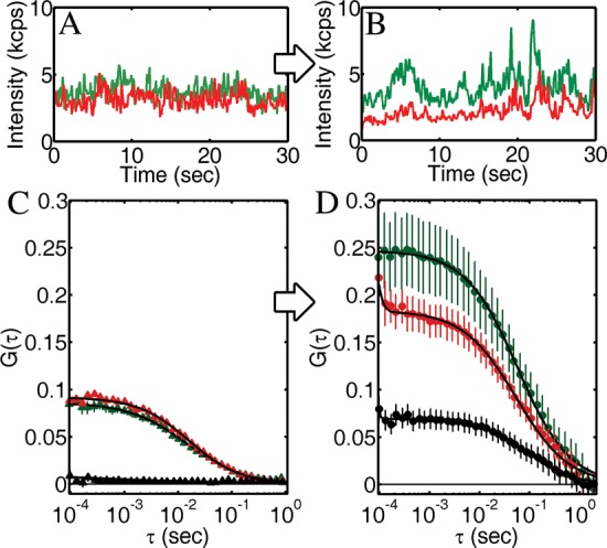
The organization and dynamics of plasma membrane components at the nanometer scale are essential for biological functions such as transmembrane signaling and endocytosis. Planarized nanoscale apertures in a metallic film are demonstrated as a means of confining the excitation light for multicolor fluorescence spectroscopy to a 55 ± 10 nm beam waist. This technique provides simultaneous two-color, subdiffraction-limited fluorescence correlation spectroscopy and fluorescence cross-correlation spectroscopy on planar membranes. The fabrication and implementation of this technique are demonstrated for both model membranes and live cells. Membrane-bound proteins were observed to cluster upon the addition of a multivalent cross-linker: On supported lipid bilayers, clusters of cholera toxin subunit B were formed upon cross-linking by an antibody specific for this protein; on living cells, immunoglobulin E bound to its receptor (FcεRI) on the plasma membranes of RBL mast cells was observed to form clusters upon exposure to a trivalent antigen. The formation of membrane clusters was quantified via fluorescence intensity vs time and changes in the temporal auto- and cross-correlations above a single nanoscale aperture. The illumination profile from a single aperture is analyzed experimentally and computationally with a rim-dominated illumination profile, yielding no change in the autocorrelation dwell time with changes in aperture diameter from 60 to 250 nm. This near-field fluorescence cross-correlation methodology provides access to nanoscale details of dynamic membrane interactions and motivates further development of near-field optical methods.
质膜成分在纳米尺度上的组织和动力学对于诸如跨膜信号传导和内吞作用等生物学功能至关重要。金属膜中的平面化纳米级孔径被证明是一种将多色荧光光谱的激发光限制在55±10纳米束腰的方法。该技术可在平面膜上同时进行双色、亚衍射极限荧光相关光谱和荧光交叉相关光谱分析。本文展示了该技术在模型膜和活细胞上的制备与应用。观察到膜结合蛋白在添加多价交联剂后会聚集:在支持的脂质双分子层上,霍乱毒素B亚基的簇在针对该蛋白的抗体交联后形成;在活细胞上,观察到RBL肥大细胞质膜上与其受体(FcεRI)结合的免疫球蛋白E在暴露于三价抗原后形成簇。通过荧光强度随时间的变化以及单个纳米级孔径上方时间自相关和交叉相关的变化对膜簇的形成进行了量化。对单个孔径的照明轮廓进行了实验和计算分析,其照明轮廓以边缘为主,随着孔径直径从60纳米变化到250纳米,自相关停留时间没有变化。这种近场荧光交叉相关方法能够获取动态膜相互作用的纳米级细节,并推动了近场光学方法的进一步发展。