Xue Ruipeng, Behera Prajna, Xu Joshua, Viapiano Mariano S, Lannutti John J
Department of Materials Science and Engineering, The Ohio State University, Columbus, OH 43210, USA.
Department of Neurosurgery, Brigham and Women's Hospital, Harvard Medical School, Boston, MA 02115, USA.
Sens Actuators B Chem. 2014 Mar 1;192:697-707. doi: 10.1016/j.snb.2013.10.084.
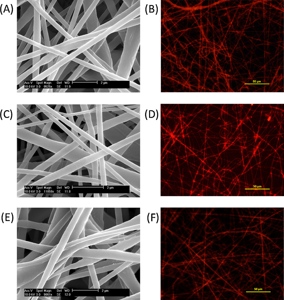
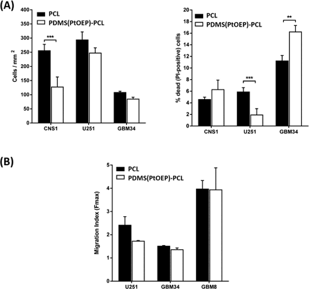
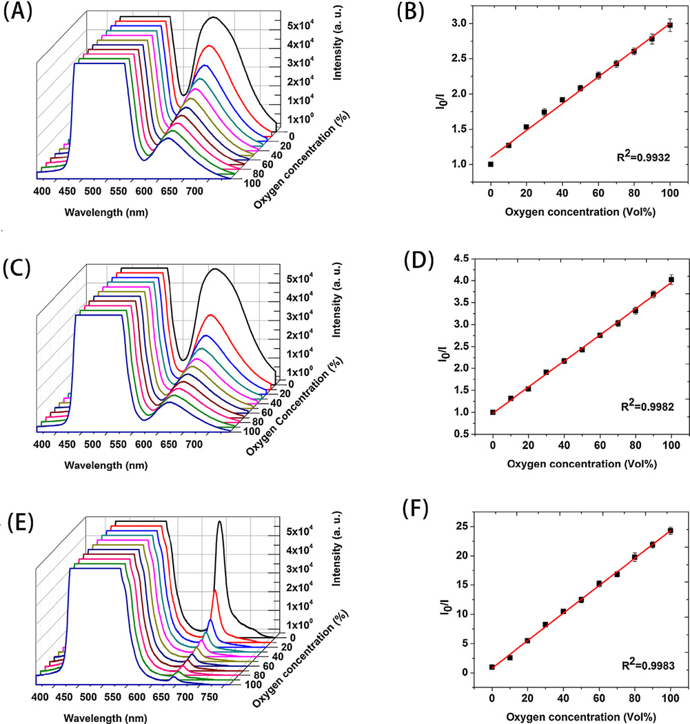
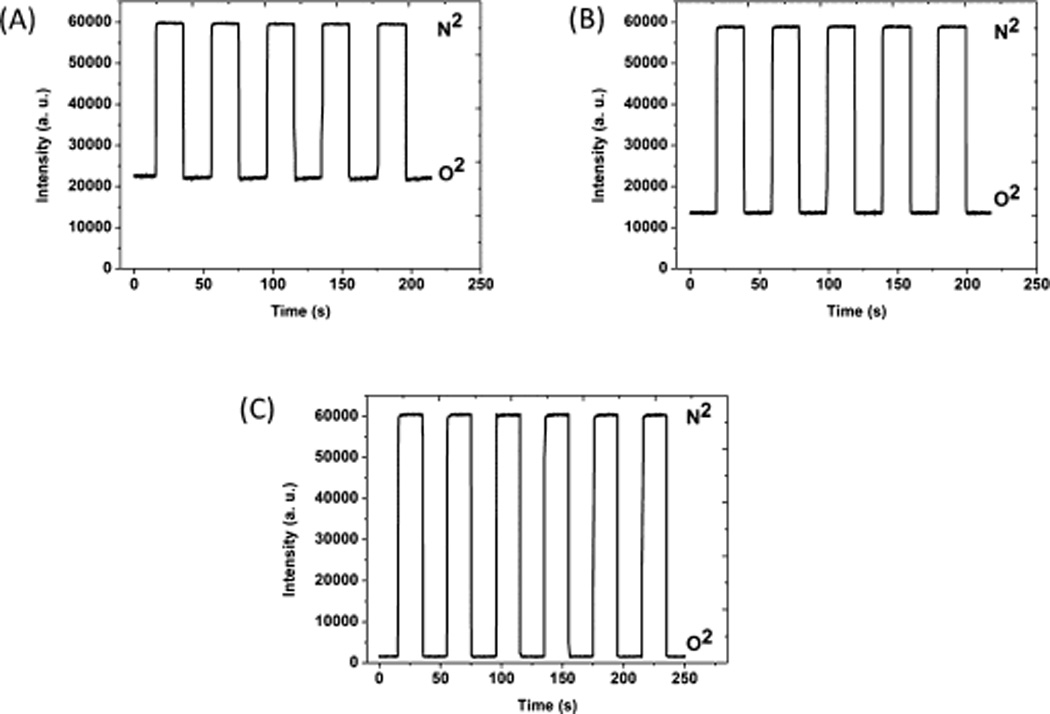
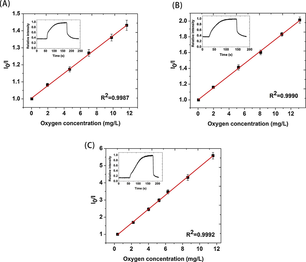
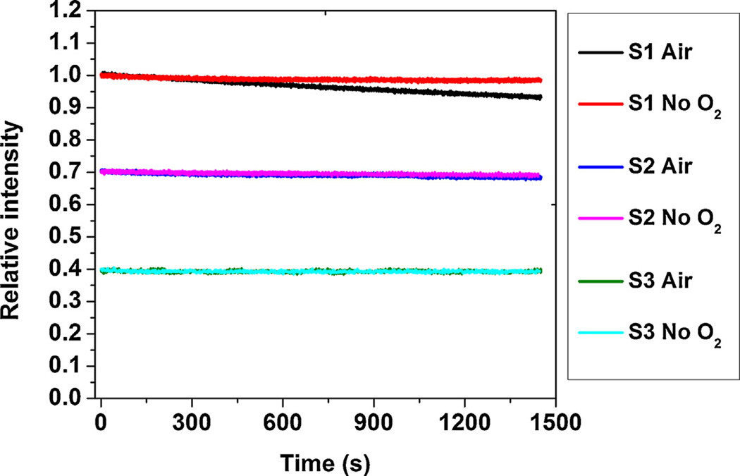
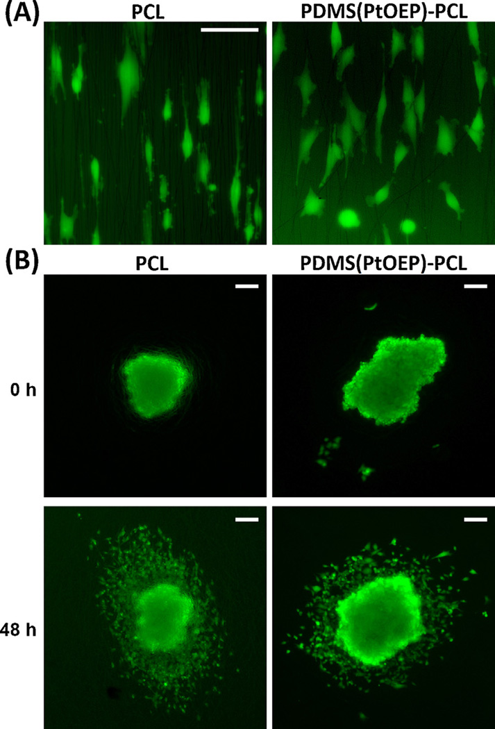
Real-time, continuous monitoring of local oxygen contents at the cellular level is desirable both for the study of cancer cell biology and in tissue engineering. In this paper, we report the successful fabrication of polydimethylsiloxane (PDMS) nanofibers containing oxygen-sensitive probes by electrospinning and the applications of these fibers as optical oxygen sensors for both gaseous and dissolved oxygen. A protective 'shell' layer of polycaprolactone (PCL) not only maintains the fiber morphology of PDMS during the slow curing process but also provides more biocompatible surfaces. Once this strategy was perfected, tris(4,7-diphenyl-1,10-phenanthroline) ruthenium(II) (Ru(dpp)) and platinum octaethylporphyrin (PtOEP) were dissolved in the PDMS core and the resulting sensing performance established. These new core-shell sensors containing different sensitivity probes showed slight variations in oxygen response but all exhibited excellent Stern-Volmer linearity. Due in part to the porous nature of the fibers and the excellent oxygen permeability of PDMS, the new sensors show faster response (<0.5 s) -4-10 times faster than previous reports - than conventional 2D film-based oxygen sensors. Such core-shell fibers are readily integrated into standard cell culture plates or bioreactors. The photostability of these nanofiber-based sensors was also assessed. Culture of glioma cell lines (CNS1, U251) and glioma-derived primary cells (GBM34) revealed negligible differences in biological behavior suggesting that the presence of the porphyrin dyes within the core carries with it no strong cytotoxic effects. The unique combination of demonstrated biocompatibility due to the PCL 'shell' and the excellent oxygen transparency of the PDMS core makes this particular sensing platform promising for sensing in the context of biological environments.
无论是对于癌细胞生物学研究还是组织工程而言,在细胞水平上对局部氧含量进行实时、连续监测都是很有必要的。在本文中,我们报告了通过静电纺丝成功制备出含有氧敏感探针的聚二甲基硅氧烷(PDMS)纳米纤维,以及这些纤维作为气态和溶解氧光学氧传感器的应用。聚己内酯(PCL)的保护性“壳”层不仅在缓慢固化过程中维持了PDMS的纤维形态,还提供了更具生物相容性的表面。一旦该策略完善,将三(4,7-二苯基-1,10-菲咯啉)钌(II)(Ru(dpp))和铂八乙基卟啉(PtOEP)溶解在PDMS核中,并建立了由此产生的传感性能。这些含有不同灵敏度探针的新型核壳传感器在氧响应方面表现出轻微差异,但均表现出优异的斯特恩-沃尔默线性。部分由于纤维的多孔性质和PDMS优异的氧渗透性,新型传感器显示出比传统二维薄膜基氧传感器更快的响应(<0.5秒)——比之前的报道快4-10倍。这种核壳纤维很容易集成到标准细胞培养板或生物反应器中。还评估了这些基于纳米纤维的传感器的光稳定性。胶质瘤细胞系(CNS1、U251)和胶质瘤衍生原代细胞(GBM34)的培养显示,生物学行为上的差异可忽略不计,这表明核内卟啉染料的存在没有强烈的细胞毒性作用。由于PCL“壳”所证明的生物相容性与PDMS核优异的氧透明度的独特结合,使得这个特殊的传感平台在生物环境传感方面很有前景。