Yoshii Hiroshi, Yanagihara Kouta, Imaseki Hitoshi, Hamano Tsuyoshi, Yamanishi Hirokuni, Inagaki Masayo, Sakai Yasuhiro, Sugiura Nobuyuki, Kurihara Osamu, Sakai Kazuo
Research Center for Radiation Emergency Medicine, National Institute of Radiological Science, Chiba, Chiba, Japan.
Research Center for Radiation Emergency Medicine, National Institute of Radiological Science, Chiba, Chiba, Japan; Department of Physics, Faculty of Science, Toho University, Funabashi, Chiba, Japan.
PLoS One. 2014 Jul 10;9(7):e101966. doi: 10.1371/journal.pone.0101966. eCollection 2014.
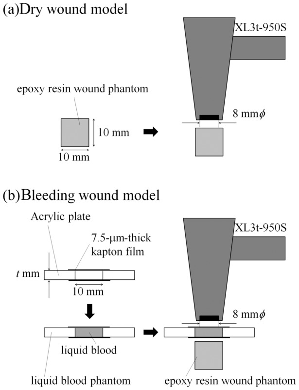
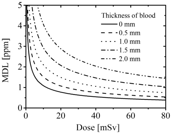
Workers decommissioning the Fukushima-Daiichi nuclear power plant damaged from the Great East Japan Earthquake and resulting tsunami are at risk of injury with possible contamination from radioactive heavy atoms including actinides, such as plutonium. We propose a new methodology for on-site and rapid evaluation of heavy-atom contamination in wounds using a portable X-ray fluorescence (XRF) device. In the present study, stable lead was used as the model contaminant substitute for radioactive heavy atoms. First, the wound model was developed by placing a liquid blood phantom on an epoxy resin wound phantom contaminated with lead. Next, the correlation between the concentration of contaminant and the XRF peak intensity was formulated considering the thickness of blood exiting the wound. Methods to determine the minimum detection limit (MDL) of contaminants at any maximal equivalent dose to the wound by XRF measurement were also established. For example, in this system, at a maximal equivalent dose of 16.5 mSv to the wound and blood thickness of 0.5 mm, the MDL value for lead was 1.2 ppm (3.1 nmol). The radioactivity of 239Pu corresponding to 3.1 nmol is 1.7 kBq, which is lower than the radioactivity of 239Pu contaminating puncture wounds in previous severe accidents. In conclusion, the established methodology could be beneficial for future development of a method to evaluate plutonium contamination in wounds. Highlights: Methodology for evaluation of heavy-atom contamination in a wound was established. A portable X-ray fluorescence device enables on-site, rapid and direct evaluation. This method is expected to be used for evaluation of plutonium contamination in wounds.
参与福岛第一核电站退役工作的工人面临受伤风险,该核电站在东日本大地震及由此引发的海啸中受损,他们可能受到包括锕系元素(如钚)在内的放射性重原子的污染。我们提出了一种新方法,使用便携式X射线荧光(XRF)设备对伤口中的重原子污染进行现场快速评估。在本研究中,稳定铅被用作放射性重原子的模型污染物替代品。首先,通过将液体血液模型放置在被铅污染的环氧树脂伤口模型上,建立伤口模型。接下来,考虑从伤口流出的血液厚度,建立污染物浓度与XRF峰值强度之间的相关性。还建立了通过XRF测量确定伤口在任何最大等效剂量下污染物最低检测限(MDL)的方法。例如,在该系统中,当伤口的最大等效剂量为16.5 mSv且血液厚度为0.5 mm时,铅的MDL值为1.2 ppm(3.1 nmol)。与3.1 nmol相对应的239Pu放射性为1.7 kBq,低于先前严重事故中污染穿刺伤口的239Pu放射性。总之,所建立的方法可能有助于未来开发评估伤口中钚污染的方法。要点:建立了评估伤口中重原子污染的方法。便携式X射线荧光设备能够进行现场、快速和直接评估。该方法有望用于评估伤口中的钚污染。