Liu Wenzhong, Schultz Kathryn M, Zhang Kevin, Sasman Amy, Gao Fengli, Kume Tsutomu, Zhang Hao F
Department of Biomedical Engineering, Northwestern University, Evanston, IL, 60208, USA.
Feinberg Cardiovascular Research Institute, Feinberg School of Medicine, Northwestern University, Chicago, IL 60611, USA.
Photoacoustics. 2014 Jun 1;2(2):81-86. doi: 10.1016/j.pacs.2014.04.003.
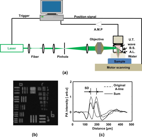
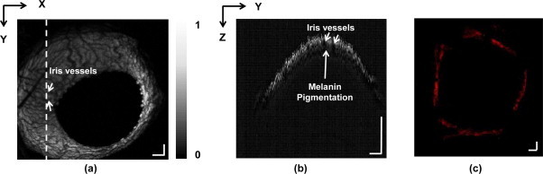
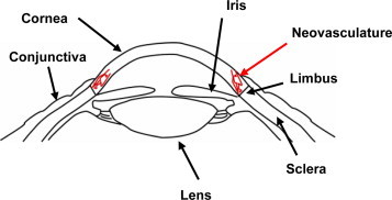
Corneal neovascularization leads to blurred vision, thus visualization is essential for pathological studies in animal models. Photoacoustic (PA) imaging can delineate microvasculature and hemodynamics noninvasively, which is suitable for investigating corneal neovascularization. In this study, we demonstrate imaging of corneal neovascularization in the mouse eye by optical-resolution photoacoustic microscopy (OR-PAM), where corneal neovascularization is induced by deliberate alkali burn injuries in C57BL6/J inbred mice corneas on the left eye. We used OR-PAM to image five mice with corneal alkali burn injuries; the uninjured eyes (right eye) in these mice are then used as the controls. Corneal images acquired by OR-PAM with and without alkali burn injury are compared, clear signs of corneal neovascularization are present in the OR-PAM images of injured eyes; the OR-PAM results are also confirmed by postmortem fluorescence-labeled confocal microscopy.
角膜新生血管化会导致视力模糊,因此可视化对于动物模型的病理学研究至关重要。光声(PA)成像可以无创地描绘微血管结构和血流动力学,适用于研究角膜新生血管化。在本研究中,我们通过光学分辨率光声显微镜(OR-PAM)展示了小鼠眼角膜新生血管化的成像,其中通过故意对C57BL6/J近交系小鼠左眼角膜进行碱烧伤来诱导角膜新生血管化。我们使用OR-PAM对五只角膜碱烧伤的小鼠进行成像;这些小鼠未受伤的眼睛(右眼)用作对照。比较了通过OR-PAM获得的有和没有碱烧伤损伤的角膜图像,受伤眼睛的OR-PAM图像中存在角膜新生血管化的明显迹象;死后荧光标记共聚焦显微镜也证实了OR-PAM的结果。