Xu Tongda, Wu Xin, Chen Qiuping, Zhu Shasha, Liu Yang, Pan Defeng, Chen Xiaohu, Li Dongye
Research Institute of Cardiovascular Diseases, Xuzhou Medical College, Xuzhou, Jiangsu, China; The First Clinical College, Nanjing Traditional Chinese Medicine University, Nanjing, Jiangsu, China.
Research Institute of Cardiovascular Diseases, Xuzhou Medical College, Xuzhou, Jiangsu, China.
PLoS One. 2014 Jul 14;9(7):e102292. doi: 10.1371/journal.pone.0102292. eCollection 2014.
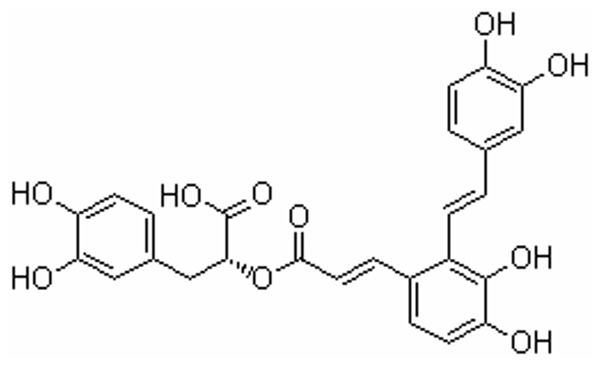
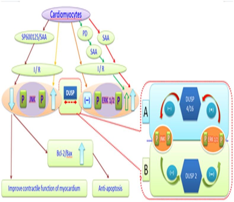
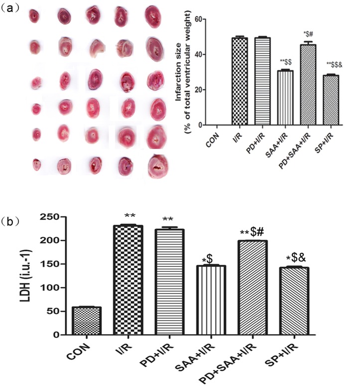
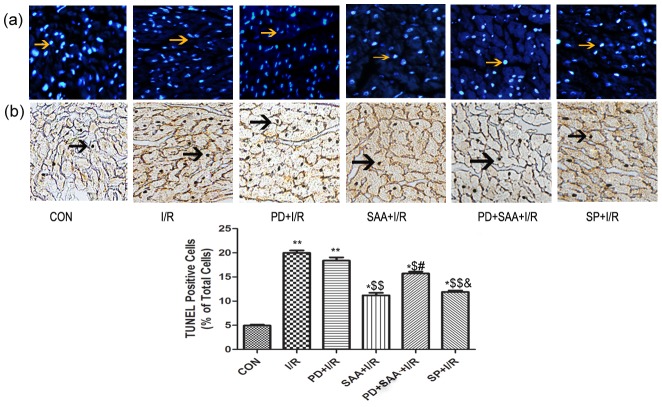
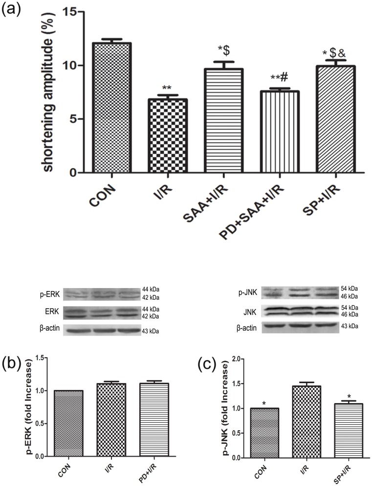
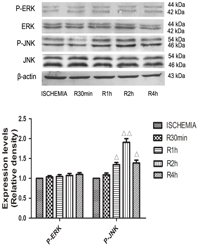
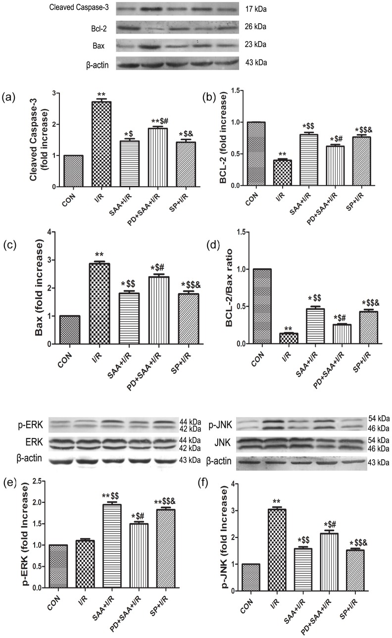
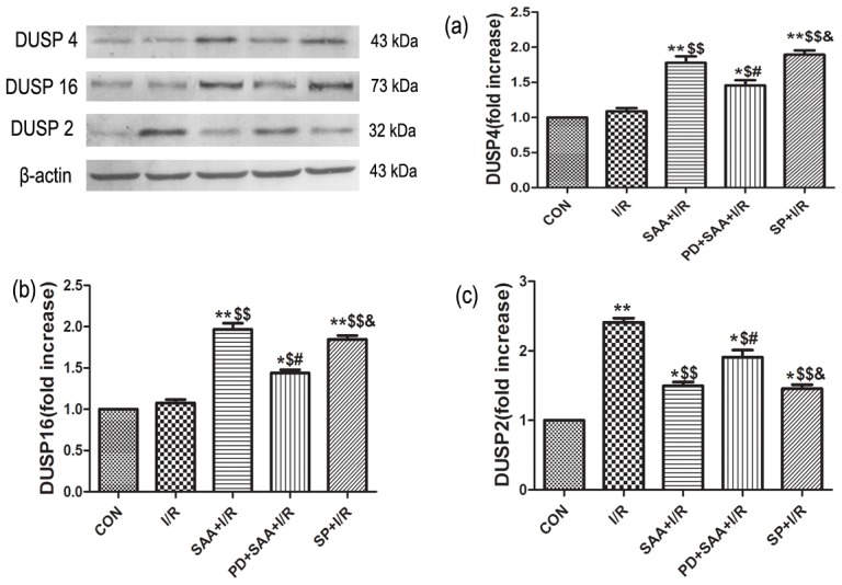
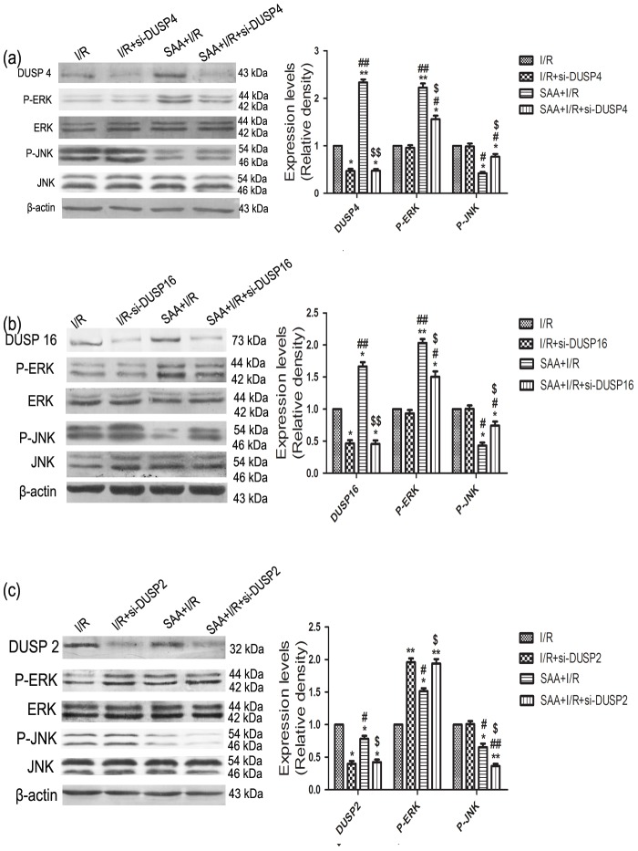
The purpose of this study was to observe the effects of salvianolic acid A (SAA) pretreatment on the myocardium during ischemia/reperfusion (I/R) and to illuminate the interrelationships among dual specificity protein phosphatase (DUSP) 2/4/16, ERK1/2 and JNK pathways during myocardial I/R, with the ultimate goal of elucidating how SAA exerts cardioprotection against I/R injury (IRI). Wistar rats were divided into the following six groups: control group (CON), I/R group, SAA+I/R group, ERK1/2 inhibitor PD098059+I/R group (PD+I/R), PD+SAA+I/R group, and JNK inhibitor SP600125+I/R group (SP+I/R). The cardioprotective effects of SAA on the myocardium during I/R were investigated with a Langendorff device. Heart rate (HR), left ventricular systolic pressure (LVSP), left ventricular end-diastolic pressure (LVEDP), maximum rate of ventricular pressure rise and fall (±dp/dtmax), myocardial infarction areas (MIA), lactate dehydrogenase (LDH), and cardiomyocytes apoptosis were monitored. To determine the crosstalk betwee JNK and ERK1/2 via DUSP2/4/16 with SAA pretreatment, siRNA-DUSP2/4/16 were performed. The expression levels of Bcl-2, Bax, caspase 3, p-JNK, p-ERK1/2 and DUSP2/4/16 in cardiomyocytes were assayed by Western blot. Our results showed that LDH, MIA and cell apoptosis were decreased, and various parameters of heart function were improved by SAA pretreatment and SP application. In the I/R group, the expression levels of p-ERK1/2 and DUSP4/16 were not significantly different compared with the CON group, however, the protein expression levels of p-ERK1/2, Bcl-2 and DUSP4/16 were higher, while p-JNK, Bax, caspase 3 and DUSP2 levels were reduced among the SAA+I/R, PD+SAA+I/R and SP+I/R groups. The above indices were not significantly different between the SAA+I/R and SP+I/R groups. Compared with the SAA+I/R group, p-ERK1/2 was increased and p-JNK was decreased in the SAA+si-DUSP2+I/R, however, p-ERK was downregulated and p-JNK was upregulated in SAA+si-DUSP4+I/R group. SAA exerts an anti-apoptotic role against myocardial IRI by inhibiting DUSP2-mediated JNK dephosphorylation and activating DUSP4/16-mediated ERK1/2 phosphorylation.
本研究旨在观察丹酚酸A(SAA)预处理对心肌缺血/再灌注(I/R)过程中心肌的影响,并阐明双特异性蛋白磷酸酶(DUSP)2/4/16、细胞外信号调节激酶1/2(ERK1/2)和应激活化蛋白激酶(JNK)通路在心肌I/R过程中的相互关系,最终目的是阐明SAA如何对I/R损伤(IRI)发挥心脏保护作用。将Wistar大鼠分为以下六组:对照组(CON)、I/R组、SAA+I/R组、ERK1/2抑制剂PD098059+I/R组(PD+I/R)、PD+SAA+I/R组和JNK抑制剂SP600125+I/R组(SP+I/R)。使用Langendorff装置研究SAA对I/R过程中心肌的心脏保护作用。监测心率(HR)、左心室收缩压(LVSP)、左心室舒张末期压力(LVEDP)、心室压力上升和下降的最大速率(±dp/dtmax)、心肌梗死面积(MIA)、乳酸脱氢酶(LDH)和心肌细胞凋亡情况。为了确定经SAA预处理后JNK和ERK1/2通过DUSP2/4/16的相互作用,进行了小干扰RNA(siRNA)-DUSP2/4/16实验。通过蛋白质免疫印迹法检测心肌细胞中Bcl-2、Bax、半胱天冬酶3、磷酸化JNK(p-JNK)、磷酸化ERK1/2(p-ERK1/2)和DUSP2/4/16的表达水平。我们的结果表明,SAA预处理和SP应用可降低LDH、MIA和细胞凋亡,并改善心功能的各项参数。在I/R组中,与CON组相比,p-ERK1/2和DUSP4/16的表达水平无显著差异,然而,在SAA+I/R、PD+SAA+I/R和SP+I/R组中,p-ERK1/2、Bcl-2和DUSP4/16的蛋白表达水平较高,而p-JNK、Bax、半胱天冬酶3和DUSP2水平降低。SAA+I/R组和SP+I/R组之间上述指标无显著差异。与SAA+I/R组相比,SAA+si-DUSP2+I/R组中p-ERK1/2升高而p-JNK降低,然而,在SAA+si-DUSP4+I/R组中p-ERK下调而p-JNK上调。SAA通过抑制DUSP2介导的JNK去磷酸化和激活DUSP4/16介导的ERK1/2磷酸化,对心肌IRI发挥抗凋亡作用。