Stratmann Mirjam, Konrad Carsten, Kugel Harald, Krug Axel, Schöning Sonja, Ohrmann Patricia, Uhlmann Christina, Postert Christian, Suslow Thomas, Heindel Walter, Arolt Volker, Kircher Tilo, Dannlowski Udo
Department of Psychiatry, University of Marburg, Marburg, Germany.
Department of Clinical Radiology, University of Münster, Münster, Germany.
PLoS One. 2014 Jul 22;9(7):e102692. doi: 10.1371/journal.pone.0102692. eCollection 2014.
Major depressive disorder is a serious psychiatric illness with a highly variable and heterogeneous clinical course. Due to the lack of consistent data from previous studies, the study of morphometric changes in major depressive disorder is still a major point of research requiring additional studies. The aim of the study presented here was to characterize and quantify regional gray matter abnormalities in a large sample of clinically well-characterized patients with major depressive disorder.
For this study one-hundred thirty two patients with major depressive disorder and 132 age- and gender-matched healthy control participants were included, 35 with their first episode and 97 with recurrent depression. To analyse gray matter abnormalities, voxel-based morphometry (VBM8) was employed on T1 weighted MRI data. We performed whole-brain analyses as well as a region-of-interest approach on the hippocampal formation, anterior cingulate cortex and amygdala, correlating the number of depressive episodes.
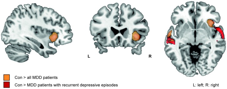
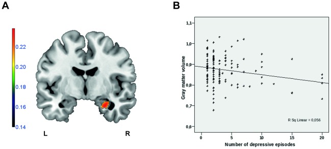
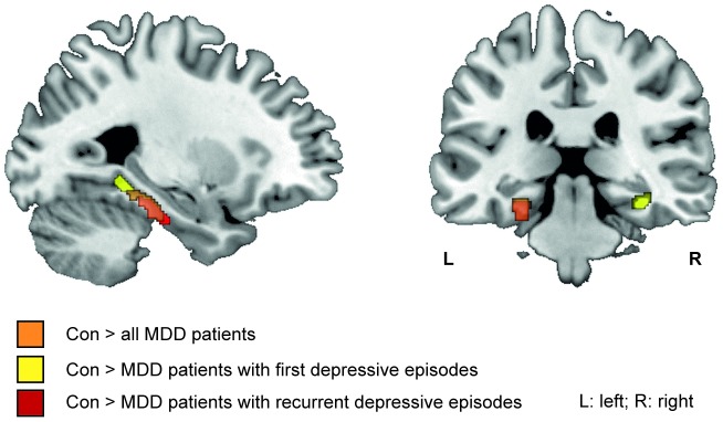
Compared to healthy control persons, patients showed a strong gray-matter reduction in the right anterior insula. In addition, region-of-interest analyses revealed significant gray-matter reductions in the hippocampal formation. The observed alterations were more severe in patients with recurrent depressive episodes than in patients with a first episode. The number of depressive episodes was negatively correlated with gray-matter volume in the right hippocampus and right amygdala.
The anterior insula gray matter structure appears to be strongly affected in major depressive disorder and might play an important role in the neurobiology of depression. The hippocampal and amygdala volume loss cumulating with the number of episodes might be explained either by repeated neurotoxic stress or alternatively by higher relapse rates in patients showing hippocampal atrophy.
重度抑郁症是一种严重的精神疾病,其临床病程高度可变且异质性强。由于先前研究缺乏一致的数据,重度抑郁症形态学变化的研究仍是需要更多研究的主要研究点。本文所述研究的目的是在大量临床特征明确的重度抑郁症患者样本中,对区域灰质异常进行表征和量化。
本研究纳入了132例重度抑郁症患者和132例年龄及性别匹配的健康对照参与者,其中35例为首次发作,97例为复发性抑郁症。为分析灰质异常,对T1加权MRI数据采用基于体素的形态学分析(VBM8)。我们进行了全脑分析以及对海马结构、前扣带回皮质和杏仁核的感兴趣区域分析,并将其与抑郁发作次数相关联。
与健康对照者相比,患者右侧前岛叶灰质明显减少。此外,感兴趣区域分析显示海马结构灰质显著减少。复发性抑郁发作患者观察到的改变比首次发作患者更严重。抑郁发作次数与右侧海马和右侧杏仁核的灰质体积呈负相关。
前岛叶灰质结构在重度抑郁症中似乎受到强烈影响,可能在抑郁症的神经生物学中起重要作用。海马和杏仁核体积损失随发作次数累积,可能是由于反复的神经毒性应激,或者是由于海马萎缩患者的较高复发率。