Zhong Yi, Utriainen David, Wang Ying, Kang Yan, Haacke E Mark
School of Sino-Dutch Biomedical and Information Engineering, Northeastern University, Shenyang, Liaoning 110004, China ; Magnetic Resonance Innovations Inc., 440 E. Ferry Street, Detroit, MI 48202, USA.
Magnetic Resonance Innovations Inc., 440 E. Ferry Street, Detroit, MI 48202, USA.
Int J Biomed Imaging. 2014;2014:239123. doi: 10.1155/2014/239123. Epub 2014 Jul 22.
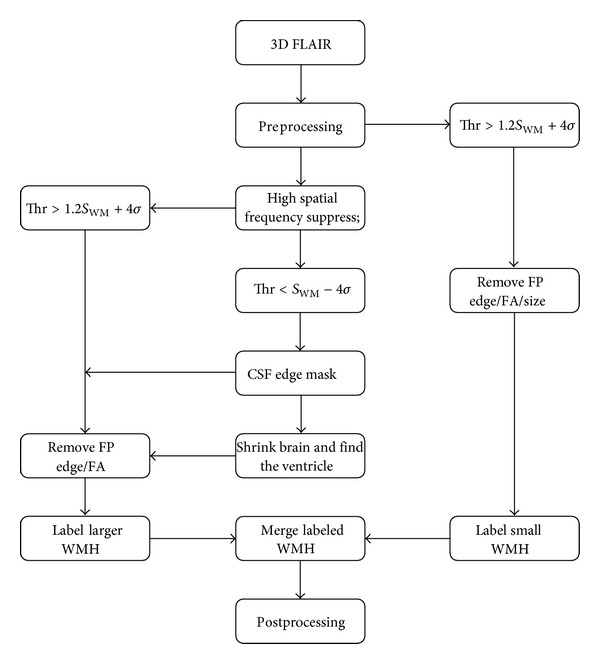
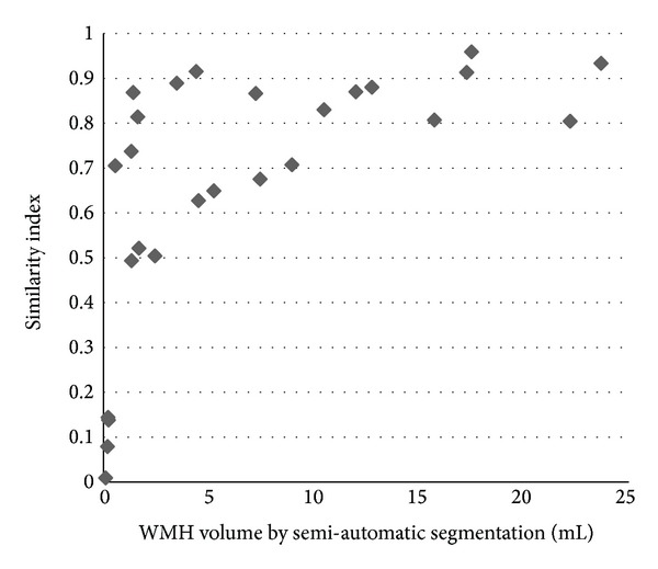
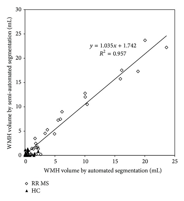
White matter hyperintensities (WMH) seen on T2WI are a hallmark of multiple sclerosis (MS) as it indicates inflammation associated with the disease. Automatic detection of the WMH can be valuable in diagnosing and monitoring of treatment effectiveness. T2 fluid attenuated inversion recovery (FLAIR) MR images provided good contrast between the lesions and other tissue; however the signal intensity of gray matter tissue was close to the lesions in FLAIR images that may cause more false positives in the segment result. We developed and evaluated a tool for automated WMH detection only using high resolution 3D T2 fluid attenuated inversion recovery (FLAIR) MR images. We use a high spatial frequency suppression method to reduce the gray matter area signal intensity. We evaluate our method in 26 MS patients and 26 age matched health controls. The data from the automated algorithm showed good agreement with that from the manual segmentation. The linear correlation between these two approaches in comparing WMH volumes was found to be Y = 1.04X + 1.74 (R (2) = 0.96). The automated algorithm estimates the number, volume, and category of WMH.
在T2加权成像(T2WI)上看到的白质高信号(WMH)是多发性硬化症(MS)的一个标志,因为它表明了与该疾病相关的炎症。WMH的自动检测在疾病诊断和治疗效果监测方面可能具有重要价值。T2液体衰减反转恢复(FLAIR)磁共振成像(MR)在病变与其他组织之间提供了良好的对比度;然而,在FLAIR图像中灰质组织的信号强度与病变接近,这可能会在分割结果中导致更多假阳性。我们开发并评估了一种仅使用高分辨率三维T2液体衰减反转恢复(FLAIR)MR图像进行WMH自动检测的工具。我们使用一种高空间频率抑制方法来降低灰质区域的信号强度。我们在26例MS患者和26例年龄匹配的健康对照中对我们的方法进行了评估。自动算法的数据与手动分割的数据显示出良好的一致性。在比较WMH体积时,发现这两种方法之间的线性相关性为Y = 1.04X + 1.74(R² = 0.96)。自动算法可估计WMH的数量、体积和类别。