Marlatt Michael W, Bauer Jan, Aronica Eleonora, van Haastert Elise S, Hoozemans Jeroen J M, Joels Marian, Lucassen Paul J
Swammerdam Institute for Life Sciences-Center for Neuroscience, University of Amsterdam, Science Park 904, 1098 XH Amsterdam, The Netherlands.
Department of Neuroimmunology, Center for Brain Research, Medical University of Vienna, Spitalgasse 4, 1090 Vienna, Austria.
Neural Plast. 2014;2014:693851. doi: 10.1155/2014/693851. Epub 2014 Aug 19.
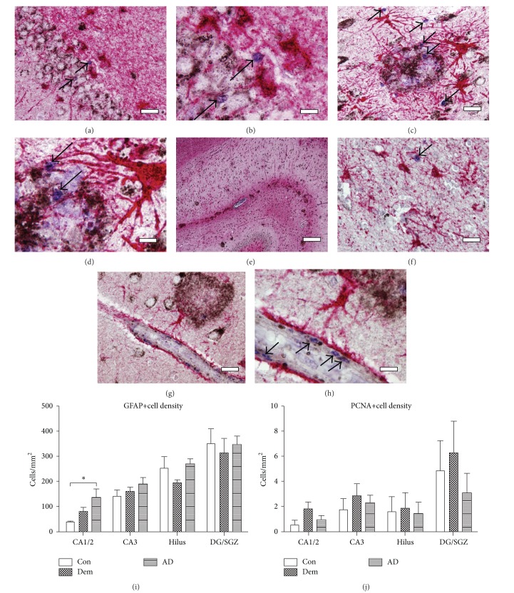
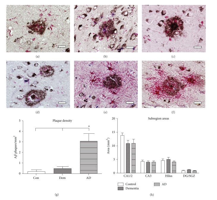
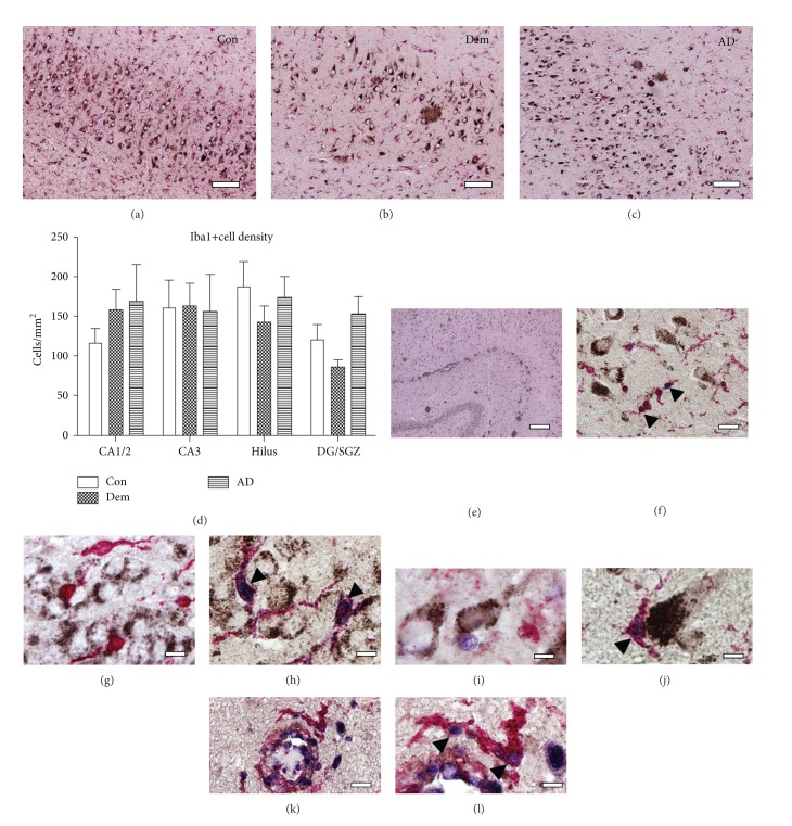
Microglia and astrocytes contribute to Alzheimer's disease (AD) etiology and may mediate early neuroinflammatory responses. Despite their possible role in disease progression and despite the fact that they can respond to amyloid deposition in model systems, little is known about whether astro- or microglia can undergo proliferation in AD and whether this is related to the clinical symptoms or to local neuropathological changes. Previously, proliferation was found to be increased in glia-rich regions of the presenile hippocampus. Since their phenotype was unknown, we here used two novel triple-immunohistochemical protocols to study proliferation in astro- or microglia in relation to amyloid pathology. We selected different age-matched cohorts to study whether proliferative changes relate to clinical severity or to neuropathological changes. Proliferating cells were found across the hippocampus but never in mature neurons or astrocytes. Almost all proliferating cells were co-labeled with Iba1+, indicating that particularly microglia contribute to proliferation in AD. Proliferating Iba1+ cells was specifically seen within the borders of amyloid plaques, indicative of an active involvement in, or response to, plaque accumulation. Thus, consistent with animal studies, proliferation in the AD hippocampus is due to microglia, occurs in close proximity of plaque pathology, and may contribute to the neuroinflammation common in AD.
小胶质细胞和星形胶质细胞在阿尔茨海默病(AD)的病因学中发挥作用,可能介导早期神经炎症反应。尽管它们在疾病进展中可能发挥作用,并且在模型系统中能够对淀粉样蛋白沉积作出反应,但对于星形胶质细胞或小胶质细胞在AD中是否会增殖,以及这种增殖是否与临床症状或局部神经病理变化相关,我们却知之甚少。此前,研究发现老年前期海马富含胶质细胞的区域增殖增加。由于这些细胞的表型未知,我们在此使用两种新型的三重免疫组织化学方法来研究星形胶质细胞或小胶质细胞的增殖与淀粉样病理的关系。我们选择了不同年龄匹配的队列来研究增殖变化是否与临床严重程度或神经病理变化相关。在整个海马中均发现了增殖细胞,但在成熟神经元或星形胶质细胞中从未发现。几乎所有增殖细胞都与Iba1+共同标记,这表明在AD中,尤其是小胶质细胞对增殖有贡献。增殖的Iba1+细胞特别见于淀粉样斑块边界内,这表明其积极参与或响应斑块积累。因此,与动物研究一致,AD海马中的增殖是由小胶质细胞引起的,发生在斑块病理附近,并且可能导致AD中常见的神经炎症。