Cipriani Paola, Di Benedetto Paola, Capece Daria, Zazzeroni Francesca, Liakouli Vasiliki, Ruscitti Piero, Pantano Ilenia, Berardicurti Onorina, Carubbi Francesco, Alesse Edoardo, Giacomelli Roberto
Department of Applied Clinical Sciences and Biotechnology, Rheumatology Unit, School of Medicine, University of L'Aquila, Delta 6 Building, Via dell'Ospedale, 67100 L'Aquila, Italy.
Department of Applied Clinical Sciences and Biotechnology, University of L'Aquila, Coppito 2, 67100 L'Aquila, Italy.
Fibrogenesis Tissue Repair. 2014 Sep 15;7:13. doi: 10.1186/1755-1536-7-13. eCollection 2014.
Systemic sclerosis (SSc) is characterized by vascular alteration and fibrosis, the former probably leading to fibrosis via the ability of both endothelial cells and pericytes to differentiate toward myofibroblast. It is well known that vascular endothelial growth factor A (VEGF-A, hereafter referred to as VEGF) may induce a profibrotic phenotype on perivascular cells. Caveolin-1 (Cav-1) is involved in the regulation of VEGF signaling, playing a role in the transport of internalized VEGF receptor 2 (VEGFR2) toward degradation, thus decreasing VEGF signaling. In this work, we assessed the levels of Cav-1 in SSc bone marrow mesenchymal stem cells (SSc-MSCs), a pericyte surrogate, and correlate these results with VEGF signaling, focusing onpotential pathogenic pathways leading to fibrosis.
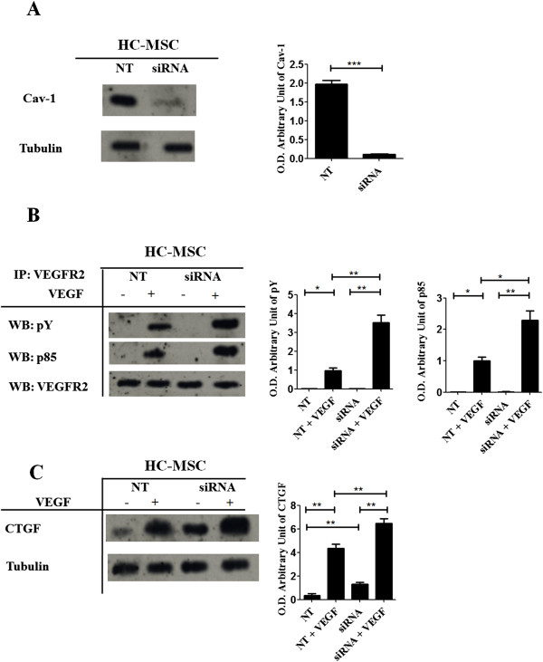
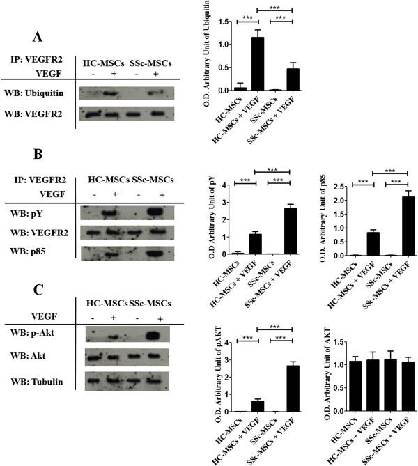
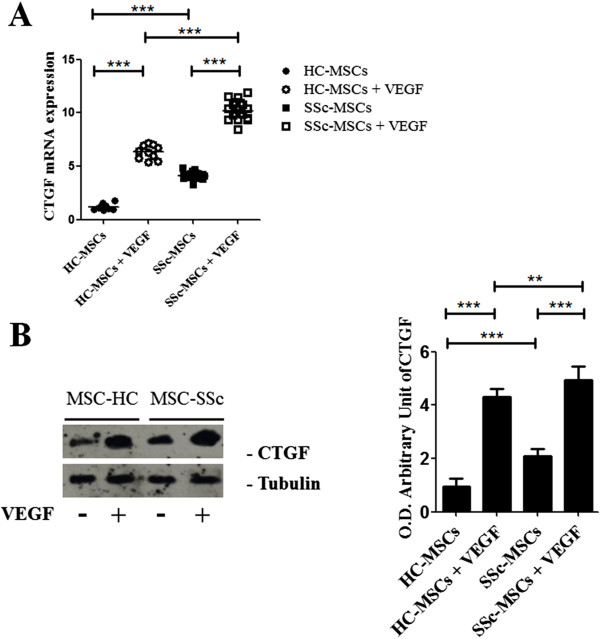
WE EXPLORED THE VEGF SIGNALING ASSESSING: (1) Cav-1 expression; (2) its co-localization with VEGFR2; (3) the activity of VEGFR2, by IF, immunoprecipitation, and western blot. In SSc-MSCs, Cav-1 levels were lower when compared to healthy controls (HC)-MSCs. Furthermore, the Cav-1/VEGFR2 co-localization and the ubiquitination of VEGFR2 were impaired in SSc-MSCs, suggesting a decreased degradation of the receptor and, as a consequence, the tyrosine phosphorylation of VEGFR2 and the PI3-kinase-Akt pathways were significantly increased when compared to HC. Furthermore, an increased connective tissue growth factor (CTGF) expression was observed in SSc-MSCs. Taken together, these data suggested the upregulation of VEGF signaling in SSc-MSCs. Furthermore, after silencing Cav-1 expression in HC-MSCs, an increased CTGF expression in HC-MSCs was observed, mirroring the results obtained in SSc-MSCs, and confirming the potential role that the lack of Cav-1 may play in the persistent VEGF signaling .
During SSc, the lower levels of Cav-1 may contribute to the pathogenesis of fibrosis via an upregulation of the VEGF signaling in perivascular cells which are shifted to a profibrotic phenotype.
系统性硬化症(SSc)的特征是血管改变和纤维化,前者可能通过内皮细胞和周细胞向肌成纤维细胞分化的能力导致纤维化。众所周知,血管内皮生长因子A(VEGF-A,以下简称VEGF)可能诱导血管周围细胞出现促纤维化表型。小窝蛋白-1(Cav-1)参与VEGF信号传导的调节,在将内化的VEGF受体2(VEGFR2)转运至降解过程中发挥作用,从而降低VEGF信号传导。在本研究中,我们评估了SSc骨髓间充质干细胞(SSc-MSCs,一种周细胞替代物)中Cav-1的水平,并将这些结果与VEGF信号传导相关联,重点关注导致纤维化的潜在致病途径。
我们通过免疫荧光(IF)、免疫沉淀和蛋白质印迹法评估VEGF信号传导,具体包括:(1)Cav-1表达;(2)其与VEGFR2的共定位;(3)VEGFR2的活性。与健康对照(HC)-MSCs相比,SSc-MSCs中Cav-1水平较低。此外,SSc-MSCs中Cav-1/VEGFR2的共定位和VEGFR2的泛素化受损,这表明受体降解减少,结果与HC相比,VEGFR2的酪氨酸磷酸化和PI3-激酶-Akt途径显著增加。此外,在SSc-MSCs中观察到结缔组织生长因子(CTGF)表达增加。综上所述,这些数据表明SSc-MSCs中VEGF信号传导上调。此外,在HC-MSCs中沉默Cav-1表达后,观察到HC-MSCs中CTGF表达增加,这与在SSc-MSCs中获得的结果相似,并证实了Cav-1缺乏可能在持续的VEGF信号传导中发挥的潜在作用。
在SSc期间,较低水平的Cav-1可能通过上调血管周围细胞中VEGF信号传导导致纤维化的发病机制,这些细胞转变为促纤维化表型。