Barrientos-Durán Antonio, Carpenter Ellen M, Zur Nieden Nicole I, Malinin Theodore I, Rodríguez-Manzaneque Juan Carlos, Zanello Laura P
Department of Biochemistry, University of California Riverside, Riverside, CA, USA ; Pfizer-University of Granada-Junta de Andalucía Centre for Genomics and Oncological Research (GENYO), Granada, Spain.
Department of Psychiatry and Biobehavioral Sciences, UCLA School of Medicine, South Los Angeles, CA, USA.
Int J Nanomedicine. 2014 Sep 9;9:4277-91. doi: 10.2147/IJN.S62538. eCollection 2014.
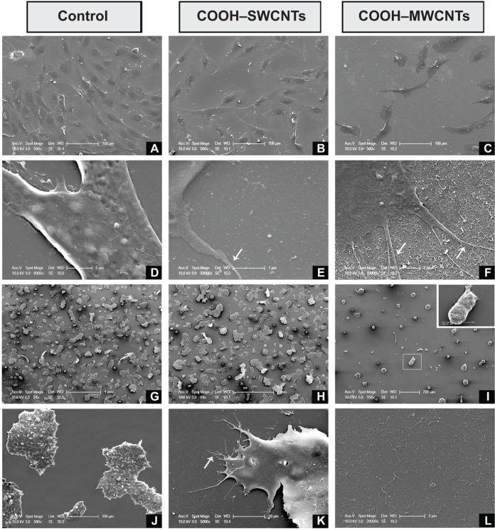
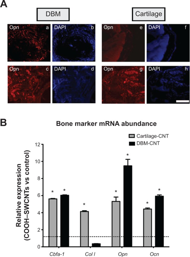
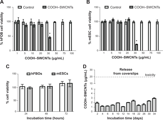
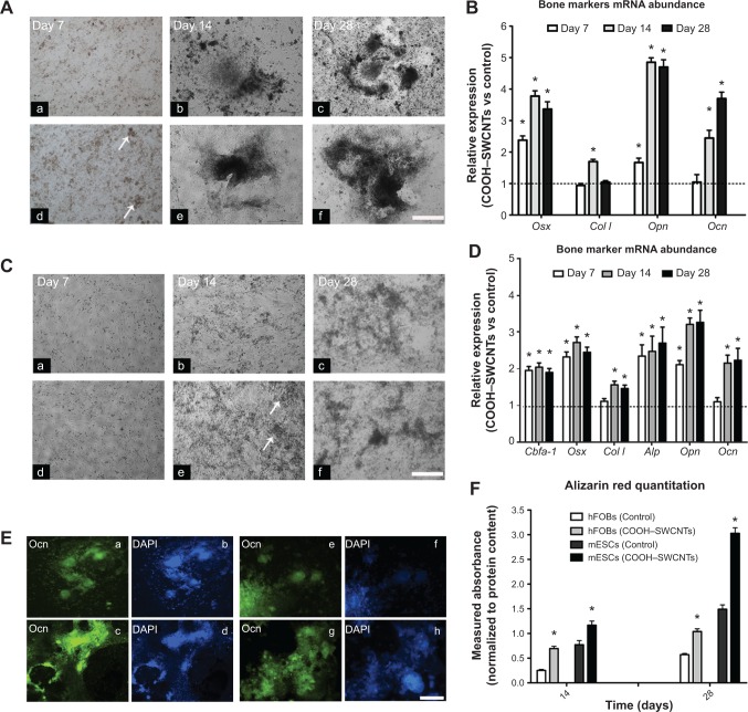
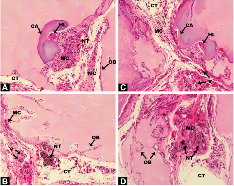
The clinical management of bone defects caused by trauma or nonunion fractures remains a challenge in orthopedic practice due to the poor integration and biocompatibility properties of the scaffold or implant material. In the current work, the osteogenic properties of carboxyl-modified single-walled carbon nanotubes (COOH-SWCNTs) were investigated in vivo and in vitro. When human preosteoblasts and murine embryonic stem cells were cultured on coverslips sprayed with COOH-SWCNTs, accelerated osteogenic differentiation was manifested by increased expression of classical bone marker genes and an increase in the secretion of osteocalcin, in addition to prior mineralization of the extracellular matrix. These results predicated COOH-SWCNTs' use to further promote osteogenic differentiation in vivo. In contrast, both cell lines had difficulties adhering to multi-walled carbon nanotube-based scaffolds, as shown by scanning electron microscopy. While a suspension of SWCNTs caused cytotoxicity in both cell lines at levels >20 μg/mL, these levels were never achieved by release from sprayed SWCNTs, warranting the approach taken. In vivo, human allografts formed by the combination of demineralized bone matrix or cartilage particles with SWCNTs were implanted into nude rats, and ectopic bone formation was analyzed. Histological analysis of both types of implants showed high permeability and pore connectivity of the carbon nanotube-soaked implants. Numerous vascularization channels appeared in the formed tissue, additional progenitor cells were recruited, and areas of de novo ossification were found 4 weeks post-implantation. Induction of the expression of bone-related genes and the presence of secreted osteopontin protein were also confirmed by quantitative polymerase chain reaction analysis and immunofluorescence, respectively. In summary, these results are in line with prior contributions that highlight the suitability of SWCNTs as scaffolds with high bone-inducing capabilities both in vitro and in vivo, confirming them as alternatives to current bone-repair therapies.
由于支架或植入材料的整合性和生物相容性较差,创伤或骨折不愈合所致骨缺损的临床管理仍是骨科实践中的一项挑战。在当前研究中,对羧基修饰的单壁碳纳米管(COOH-SWCNTs)的成骨特性进行了体内和体外研究。当人原代成骨细胞和小鼠胚胎干细胞在喷涂有COOH-SWCNTs的盖玻片上培养时,除细胞外基质提前矿化外,经典骨标志物基因表达增加以及骨钙素分泌增多表明成骨分化加速。这些结果预示着COOH-SWCNTs可用于在体内进一步促进成骨分化。相比之下,扫描电子显微镜显示,两种细胞系均难以附着于多壁碳纳米管基支架。虽然SWCNTs悬浮液在浓度>20μg/mL时对两种细胞系均具有细胞毒性,但喷涂的SWCNTs释放量从未达到该水平,因此该方法可行。在体内,将由脱矿骨基质或软骨颗粒与SWCNTs组合形成的人同种异体移植物植入裸鼠体内,并分析异位骨形成情况。对两种类型植入物的组织学分析显示,碳纳米管浸泡的植入物具有高渗透性和孔隙连通性。在形成的组织中出现了许多血管化通道,募集了更多的祖细胞,并且在植入后4周发现了新生骨化区域。定量聚合酶链反应分析和免疫荧光分别证实了骨相关基因表达的诱导以及分泌的骨桥蛋白的存在。总之,这些结果与之前的研究结果一致,这些研究强调了SWCNTs作为在体外和体内均具有高骨诱导能力的支架的适用性,证实它们可作为当前骨修复治疗的替代方法。