Das Kinsuk, Madhusoodan A P, Mili Bhabesh, Kumar Ajay, Saxena A C, Kumar Kuldeep, Sarkar Mihir, Singh Praveen, Srivastava Sameer, Bag Sadhan
Division of Physiology and Climatology.
Biochemistry and Food Science Section.
Int J Nanomedicine. 2017 Apr 19;12:3235-3252. doi: 10.2147/IJN.S122945. eCollection 2017.
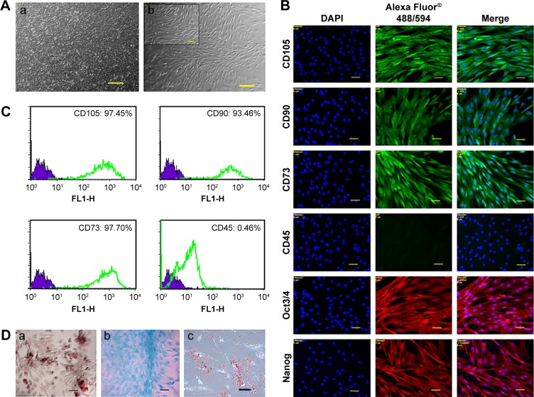
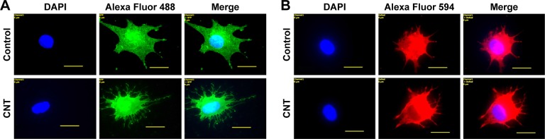
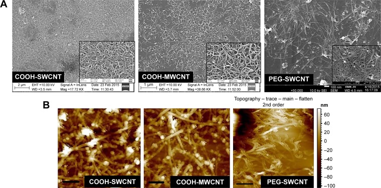
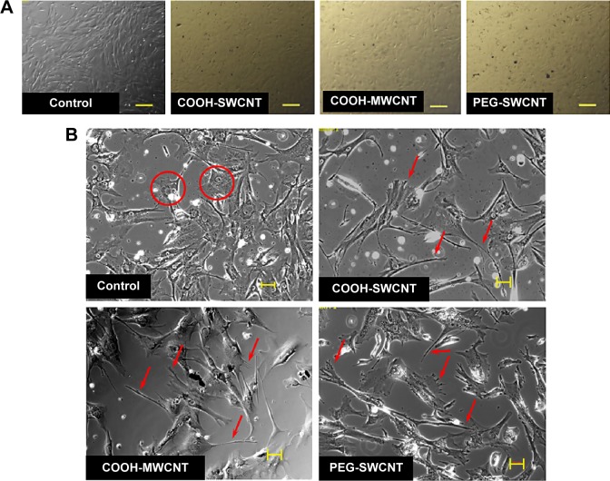
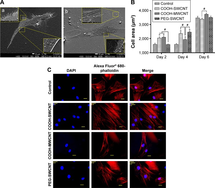
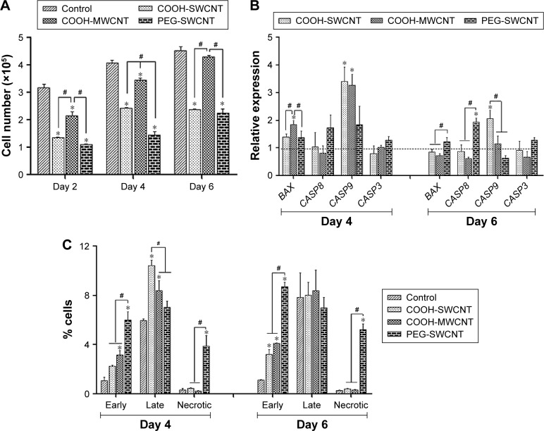
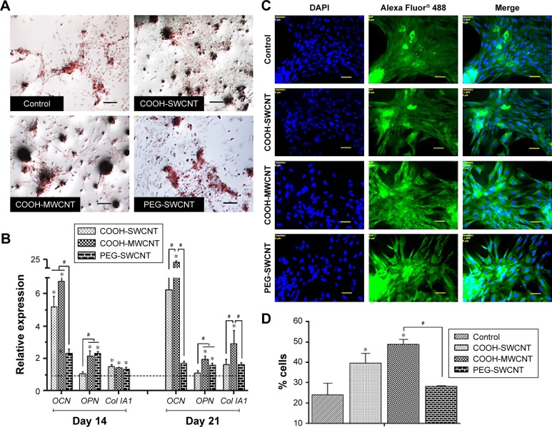
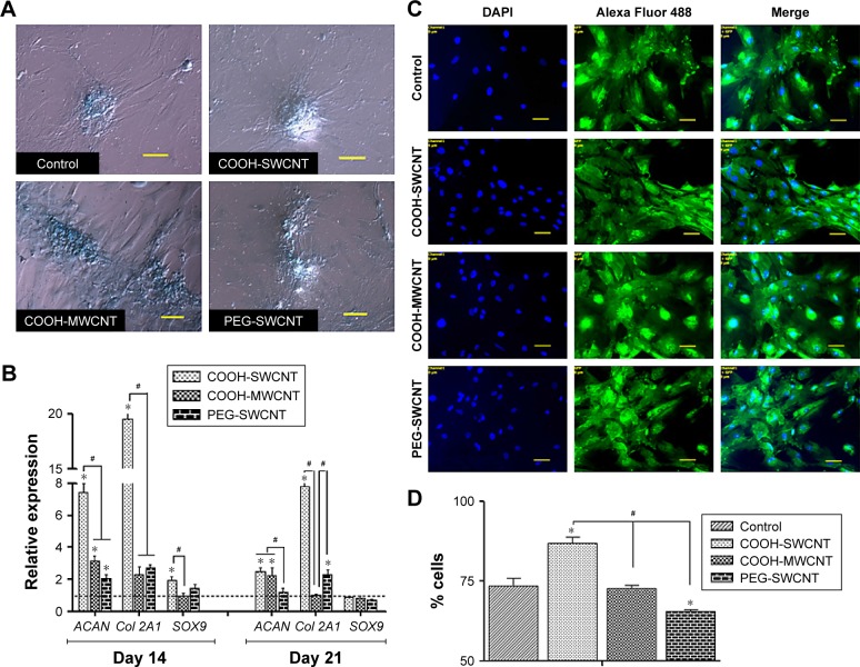
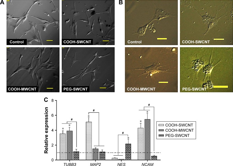
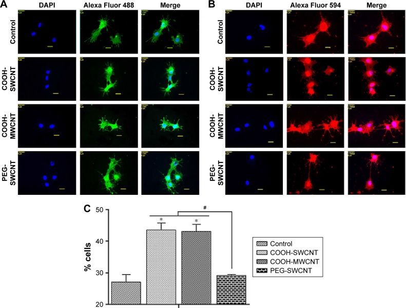
In the field of regenerative medicine, numerous potential applications of mesenchymal stem cells (MSCs) can be envisaged, due to their ability to differentiate into a range of tissues on the basis of the substrate on which they grow. With the advances in nanotechnology, carbon nanotubes (CNTs) have been widely explored for use as cell culture substrate in tissue engineering applications. In this study, canine bone marrow-derived MSCs were considered as the cellular model for an in vitro study to elucidate the collective cellular processes, using three different varieties of thin films of functionalized carbon nanotubes (COOH-single-walled CNTs [SWCNTs], COOH-multiwalled CNTs [MWCNTs] and polyethylene glycol [PEG]-SWCNTs), which were spray dried onto preheated cover slips. Cells spread out better on the CNT films, resulting in higher cell surface area and occurrence of filopodia, with parallel orientation of stress fiber bundles. Canine MSCs proliferated at a slower rate on all types of CNT substrates compared to the control, but no decline in cell number was noticed during the study period. Expression of apoptosis-associated genes decreased on the CNT substrates as time progressed. On flow cytometry after AnnexinV-fluorescein isothiocyanate/propidium iodide (PI) staining, total number of apoptotic and necrotic cells remained lower in COOH-functionalized films compared to PEG-functionalized ones. Collectively, these results indicate that COOH-MWCNT substrate provided an environment of low cytotoxicity. Canine MSCs were further induced to differentiate along osteogenic, chondrogenic, and neuronal lineages by culturing under specific differentiation conditions. The cytochemical and immunocytochemical staining results, as well as the expression of the bone marker genes, led us to hypothesize that the COOH-MWCNT substrate acted as a better cue, accelerating the osteogenic differentiation process. However, while chondrogenesis was promoted by COOH-SWCNT, neuronal differentiation was promoted by both COOH-SWNCT and COOH-MWCNT. Taken together, these findings suggest that COOH-functionalized CNTs represent a promising scaffold component for future utilization in the selective differentiation of canine MSCs in regenerative medicine.
在再生医学领域,由于间充质干细胞(MSCs)能够根据其生长的底物分化为多种组织,因此可以设想其具有众多潜在应用。随着纳米技术的进步,碳纳米管(CNTs)已被广泛探索用作组织工程应用中的细胞培养底物。在本研究中,犬骨髓来源的间充质干细胞被用作体外研究的细胞模型,以阐明集体细胞过程,使用三种不同类型的功能化碳纳米管薄膜(羧基化单壁碳纳米管[SWCNTs]、羧基化多壁碳纳米管[MWCNTs]和聚乙二醇[PEG]-SWCNTs),这些薄膜通过喷雾干燥到预热的盖玻片上。细胞在碳纳米管薄膜上铺展得更好,导致细胞表面积更大且出现丝状伪足,应力纤维束呈平行排列。与对照组相比,犬间充质干细胞在所有类型的碳纳米管底物上的增殖速率较慢,但在研究期间未观察到细胞数量下降。随着时间的推移,碳纳米管底物上凋亡相关基因的表达下降。在AnnexinV-异硫氰酸荧光素/碘化丙啶(PI)染色后的流式细胞术中,与PEG功能化薄膜相比,羧基化功能化薄膜中凋亡和坏死细胞的总数仍然较低。总体而言,这些结果表明羧基化多壁碳纳米管底物提供了低细胞毒性的环境。通过在特定分化条件下培养,犬间充质干细胞进一步被诱导沿成骨、软骨生成和神经谱系分化。细胞化学和免疫细胞化学染色结果以及骨标记基因的表达使我们推测羧基化多壁碳纳米管底物起到了更好的引导作用,加速了成骨分化过程。然而,虽然羧基化单壁碳纳米管促进软骨生成,羧基化单壁碳纳米管和羧基化多壁碳纳米管都促进神经分化。综上所述,这些发现表明羧基化功能化碳纳米管是再生医学中未来用于犬间充质干细胞选择性分化的有前景的支架成分。