Ho Ming-Hua, Liao Mei-Hsiu, Lin Yi-Ling, Lai Chien-Hao, Lin Pei-I, Chen Ruei-Ming
Department of Chemical Engineering, National Taiwan University of Science and Technology, Taipei, Taiwan ; Cell Physiology and Molecular Image Research Center and Department of Anesthesiology, Wan Fang Hospital, Taipei, Taiwan.
Graduate Institute of Medical Sciences, Taipei Medical University, Taipei, Taiwan.
Int J Nanomedicine. 2014 Sep 9;9:4293-304. doi: 10.2147/IJN.S68012. eCollection 2014.
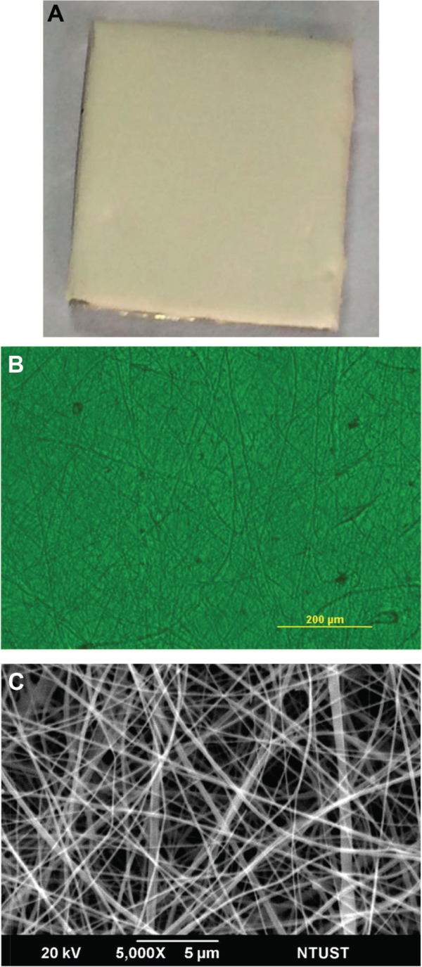
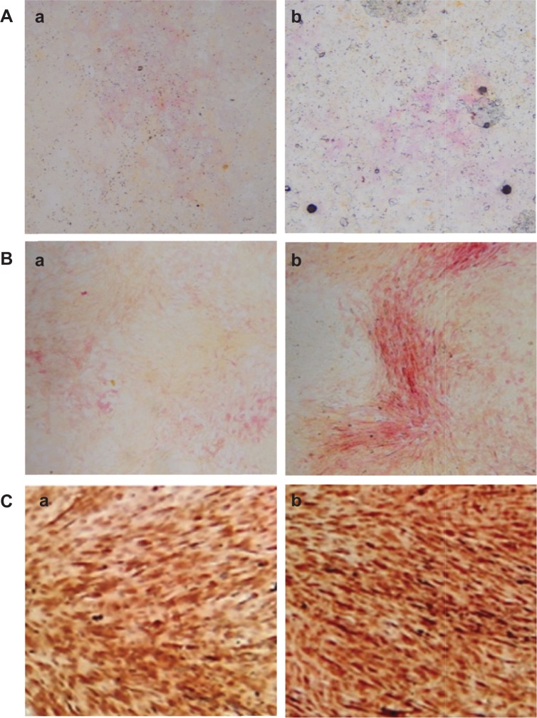
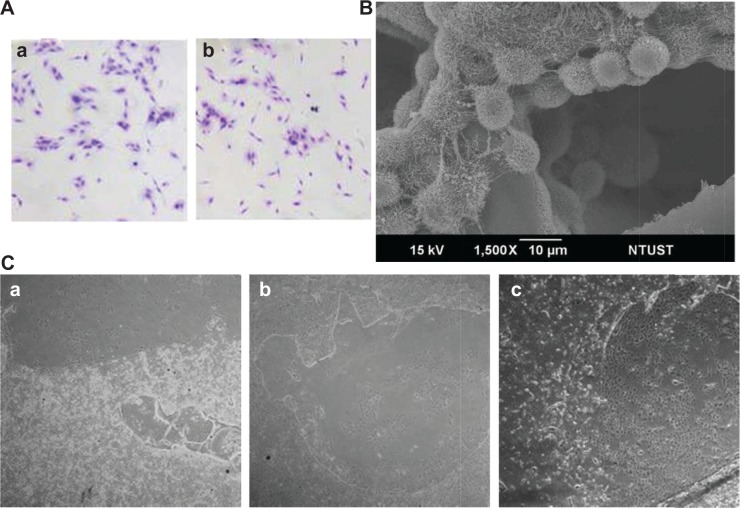
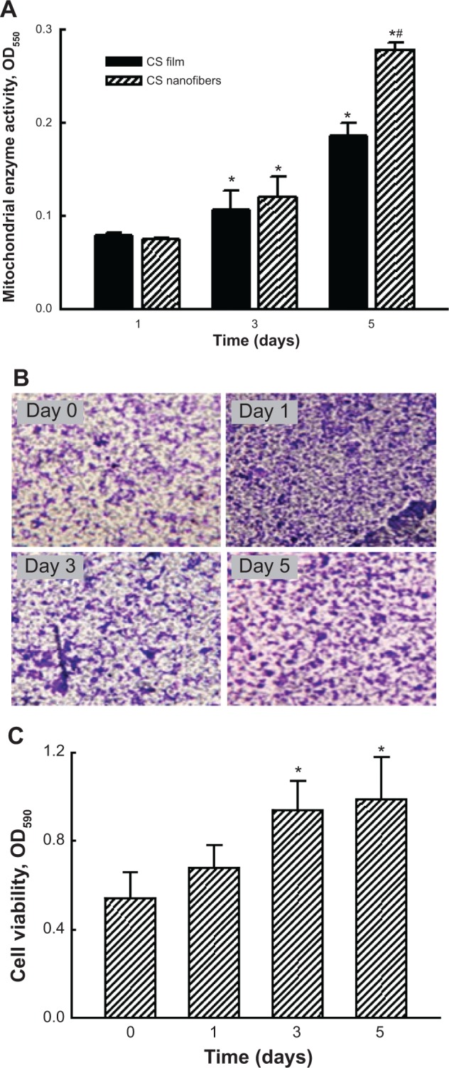
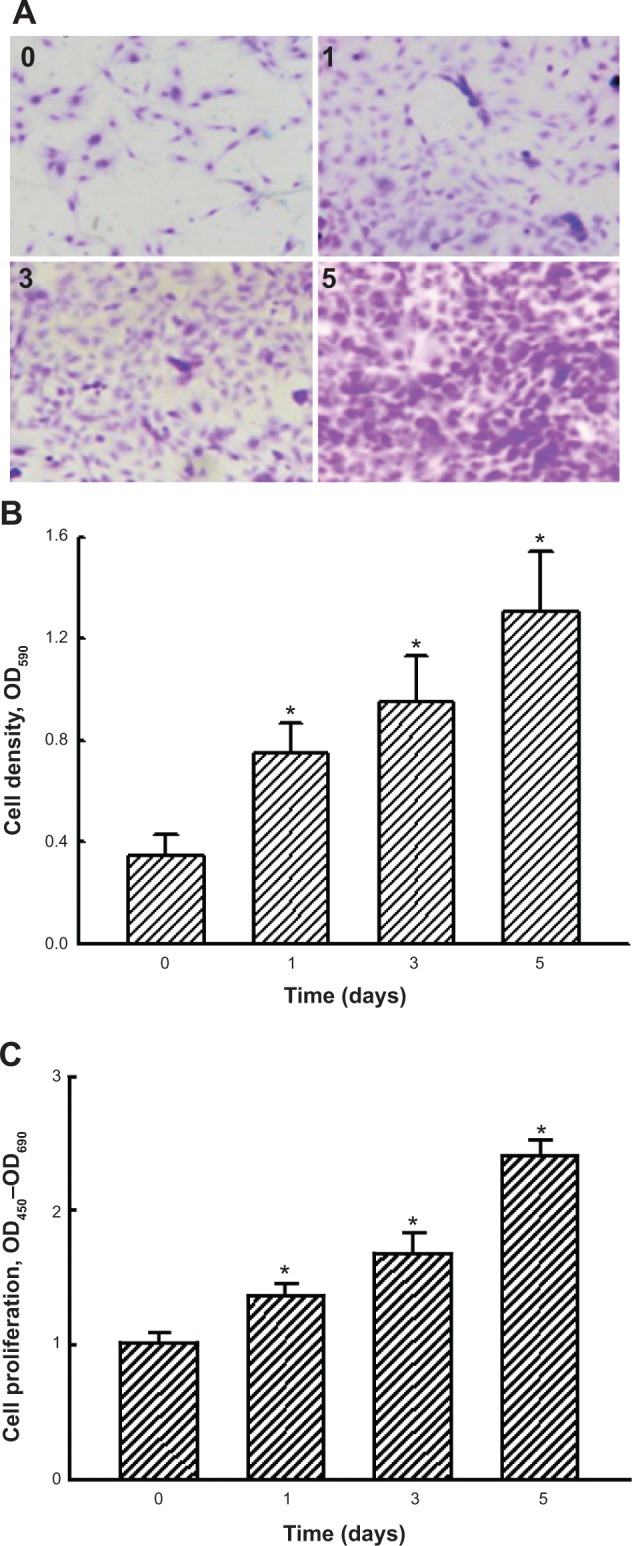
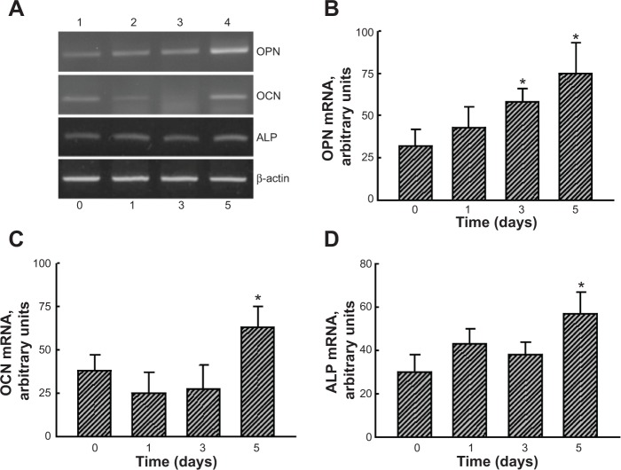
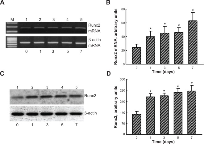
Osteoblast maturation plays a key role in regulating osteogenesis. Electrospun nanofibrous products were reported to possess a high surface area and porosity. In this study, we developed chitosan nanofibers and examined the effects of nanofibrous scaffolds on osteoblast maturation and the possible mechanisms. Macro- and micro observations of the chitosan nanofibers revealed that these nanoproducts had a flat surface and well-distributed fibers with nanoscale diameters. Mouse osteoblasts were able to attach onto the chitosan nanofiber scaffolds, and the scaffolds degraded in a time-dependent manner. Analysis by scanning electron microscopy further showed mouse osteoblasts adhered onto the scaffolds along the nanofibers, and cell-cell communication was also detected. Mouse osteoblasts grew much better on chitosan nanofiber scaffolds than on chitosan films. In addition, human osteoblasts were able to adhere and grow on the chitosan nanofiber scaffolds. Interestingly, culturing human osteoblasts on chitosan nanofiber scaffolds time-dependently increased DNA replication and cell proliferation. In parallel, administration of human osteoblasts onto chitosan nanofibers significantly induced osteopontin, osteocalcin, and alkaline phosphatase (ALP) messenger (m)RNA expression. As to the mechanism, chitosan nanofibers triggered runt-related transcription factor 2 mRNA and protein syntheses. Consequently, results of ALP-, alizarin red-, and von Kossa-staining analyses showed that chitosan nanofibers improved osteoblast mineralization. Taken together, results of this study demonstrate that chitosan nanofibers can stimulate osteoblast proliferation and maturation via runt-related transcription factor 2-mediated regulation of osteoblast-associated osteopontin, osteocalcin, and ALP gene expression.
成骨细胞成熟在调节骨生成中起关键作用。据报道,电纺纳米纤维产品具有高表面积和孔隙率。在本研究中,我们制备了壳聚糖纳米纤维,并研究了纳米纤维支架对成骨细胞成熟的影响及其可能的机制。对壳聚糖纳米纤维的宏观和微观观察表明,这些纳米产品表面平整,纤维分布均匀,直径为纳米级。小鼠成骨细胞能够附着在壳聚糖纳米纤维支架上,并且支架以时间依赖性方式降解。扫描电子显微镜分析进一步显示,小鼠成骨细胞沿纳米纤维附着在支架上,并且还检测到细胞间通讯。小鼠成骨细胞在壳聚糖纳米纤维支架上比在壳聚糖膜上生长得更好。此外,人成骨细胞能够在壳聚糖纳米纤维支架上附着和生长。有趣的是,在壳聚糖纳米纤维支架上培养人成骨细胞可使DNA复制和细胞增殖随时间增加。同时,将人成骨细胞接种到壳聚糖纳米纤维上可显著诱导骨桥蛋白、骨钙素和碱性磷酸酶(ALP)信使(m)RNA表达。至于机制,壳聚糖纳米纤维触发了与 runt 相关的转录因子 2 mRNA 和蛋白质合成。因此,ALP、茜素红和冯·科萨染色分析结果表明,壳聚糖纳米纤维改善了成骨细胞矿化。综上所述,本研究结果表明,壳聚糖纳米纤维可通过与 runt 相关的转录因子 2 介导的对成骨细胞相关的骨桥蛋白、骨钙素和 ALP 基因表达的调节来刺激成骨细胞增殖和成熟。