Böhme Steffi, Stärk Hans-Joachim, Meißner Tobias, Springer Armin, Reemtsma Thorsten, Kühnel Dana, Busch Wibke
Department of Bioanalytical Ecotoxicology, Helmholtz Centre for Environmental Research - UFZ, Permoserstr. 15, 04318 Leipzig, Germany.
Department of Analytical Chemistry, Helmholtz Centre for Environmental Research - UFZ, Permoserstr. 15, 04318 Leipzig, Germany.
J Nanopart Res. 2014;16(9):2592. doi: 10.1007/s11051-014-2592-y. Epub 2014 Aug 8.
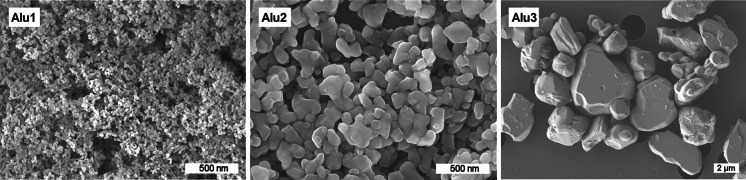
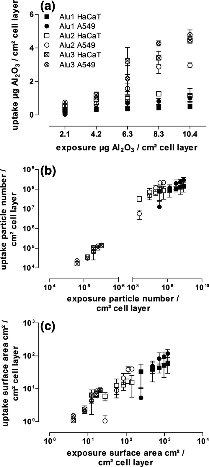
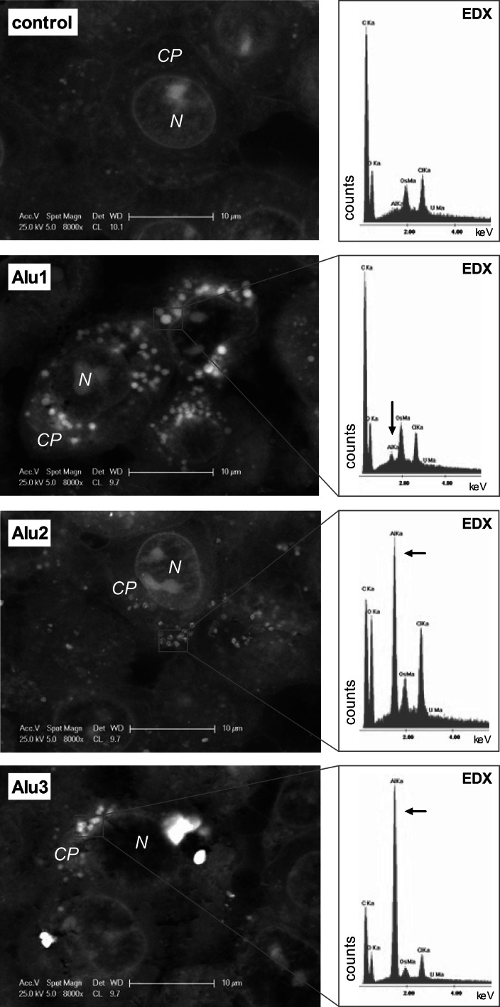
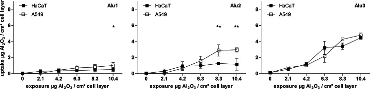
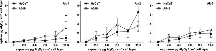
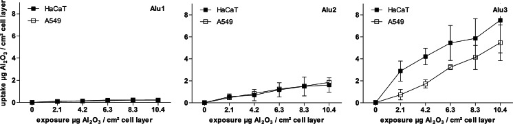
In order to quantify and compare the uptake of aluminum oxide nanoparticles of three different sizes into two human cell lines (skin keratinocytes (HaCaT) and lung epithelial cells (A549)), three analytical methods were applied: digestion followed by nebulization inductively coupled plasma mass spectrometry (neb-ICP-MS), direct laser ablation ICP-MS (LA-ICP-MS), and flow cytometry. Light and electron microscopy revealed an accumulation and agglomeration of all particle types within the cell cytoplasm, whereas no particles were detected in the cell nuclei. The internalized AlO particles exerted no toxicity in the two cell lines after 24 h of exposure. The smallest particles with a primary particle size () of 14 nm (Alu1) showed the lowest sedimentation velocity within the cell culture media, but were calculated to have settled completely after 20 h. Alu2 ( = 111 nm) and Alu3 ( = 750 nm) were calculated to reach the cell surface after 7 h and 3 min, respectively. The internal concentrations determined with the different methods lay in a comparable range of 2-8 µg AlO/cm cell layer, indicating the suitability of all methods to quantify the nanoparticle uptake. Nevertheless, particle size limitations of analytical methods using optical devices were demonstrated for LA-ICP-MS and flow cytometry. Furthermore, the consideration and comparison of particle properties as parameters for particle internalization revealed the particle size and the exposure concentration as determining factors for particle uptake.
为了量化和比较三种不同尺寸的氧化铝纳米颗粒被两种人类细胞系(皮肤角质形成细胞(HaCaT)和肺上皮细胞(A549))摄取的情况,应用了三种分析方法:消解后雾化电感耦合等离子体质谱法(neb-ICP-MS)、直接激光烧蚀ICP-MS(LA-ICP-MS)和流式细胞术。光学显微镜和电子显微镜显示所有颗粒类型在细胞质内均有积累和团聚,而在细胞核中未检测到颗粒。暴露24小时后,内化的AlO颗粒在两种细胞系中均未表现出毒性。初级粒径()为14 nm的最小颗粒(Alu1)在细胞培养基中的沉降速度最低,但经计算在20小时后已完全沉降。Alu2( = 111 nm)和Alu3( = 750 nm)经计算分别在7小时和3分钟后到达细胞表面。用不同方法测定的内部浓度在2 - 8 μg AlO/cm细胞层的可比范围内,表明所有方法都适用于量化纳米颗粒的摄取。然而,LA-ICP-MS和流式细胞术证明了使用光学设备的分析方法存在粒径限制。此外,将颗粒性质作为颗粒内化参数进行考量和比较后发现,粒径和暴露浓度是颗粒摄取的决定因素。