Hoopes P Jack, Petryk Alicia A, Gimi Barjor, Giustini Andrew J, Weaver John B, Bischof John, Chamberlain Ryan, Garwood Michael
Thayer School of Engineering at Dartmouth, 8000 Cummings Hall, Hanover, NH, USA 03755 ; Dartmouth Medical School (Departments of Surgery, Radiation Oncology and Radiology), 1 Rope Ferry Rd., Hanover, NH, USA 03755.
Thayer School of Engineering at Dartmouth, 8000 Cummings Hall, Hanover, NH, USA 03755.
Proc SPIE Int Soc Opt Eng. 2012 Mar 23;8317. doi: 10.1117/12.916097.
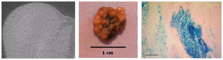
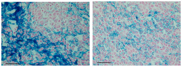
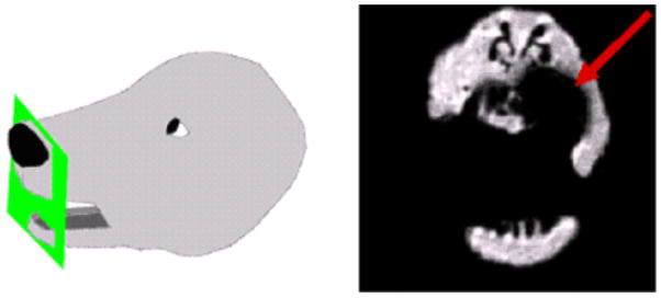
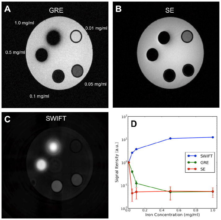
Recent advances in nanotechnology have allowed for the effective use of iron oxide nanoparticles (IONPs) for cancer imaging and therapy. When activated by an alternating magnetic field (AMF), intra-tumoral IONPs have been effective at controlling tumor growth in rodent models. To accurately plan and assess IONP-based therapies in clinical patients, noninvasive and quantitative imaging technique for the assessment of IONP uptake and biodistribution will be necessary. Proven techniques such as confocal, light and electron microscopy, histochemical iron staining, ICP-MS, fluorescent labeled mNPs and magnetic spectroscopy of Brownian motion (MSB), are being used to assess and quantify IONPs and in tissues. However, a proven noninvasive IONP imaging technique has not yet been developed. In this study we have demonstrated the shortcomings of computed tomography (CT) and magnetic resonance imaging (MRI) for effectively observing and quantifying iron/IONP concentrations in the clinical setting. Despite the poor outcomes of CT and standard MR sequences in the therapeutic concentration range, ultra-short T2 MRI methods such as, (SWIFT), provide a positive iron contrast enhancement and a reduced signal to noise ratio. Ongoing software development and phantom and studies, will further optimize this technique, providing accurate, clinically-relevant IONP biodistribution information.
纳米技术的最新进展使得氧化铁纳米颗粒(IONPs)能够有效地用于癌症成像和治疗。当被交变磁场(AMF)激活时,肿瘤内的IONPs在啮齿动物模型中已有效地控制了肿瘤生长。为了在临床患者中准确规划和评估基于IONP的治疗方法,需要用于评估IONP摄取和生物分布的非侵入性定量成像技术。诸如共聚焦、光学和电子显微镜、组织化学铁染色、电感耦合等离子体质谱、荧光标记的磁性纳米颗粒以及布朗运动磁谱(MSB)等成熟技术,正被用于评估和量化组织中的IONPs。然而,尚未开发出一种成熟的非侵入性IONP成像技术。在本研究中,我们已经证明了计算机断层扫描(CT)和磁共振成像(MRI)在临床环境中有效观察和量化铁/IONP浓度方面的不足。尽管CT和标准MR序列在治疗浓度范围内效果不佳,但诸如(SWIFT)等超短T2 MRI方法可提供正性铁对比增强和降低的信噪比。正在进行的软件开发以及体模和研究,将进一步优化该技术,提供准确的、与临床相关的IONP生物分布信息。