Al-Mashhadi Sufana, Simpson Julie E, Heath Paul R, Dickman Mark, Forster Gillian, Matthews Fiona E, Brayne Carol, Ince Paul G, Wharton Stephen B
Sheffield Institute for Translational Neuroscience, University of Sheffield, Sheffield, UK.
King Fahad Medical City, Riyadh, Saudi Arabia.
Brain Pathol. 2015 Sep;25(5):565-74. doi: 10.1111/bpa.12216. Epub 2014 Nov 20.
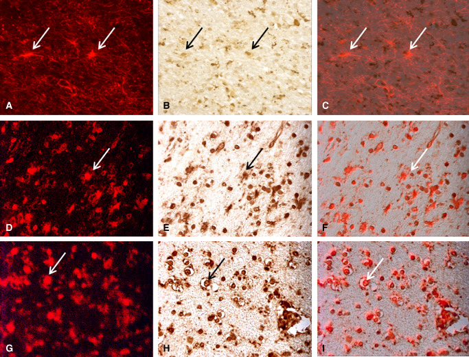
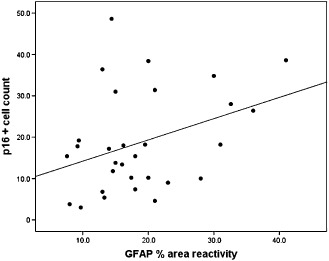
White matter lesions (WML) are common in brain aging and are associated with dementia. We aimed to investigate whether oxidative DNA damage and occur in WML and in apparently normal white matter in cases with lesions. Tissue from WML and control white matter from brains with lesions (controls lesional) and without lesions (controls non-lesional) were obtained, using post-mortem magnetic resonance imaging-guided sampling, from the Medical Research Council Cognitive Function and Ageing Study. Oxidative damage was assessed by immunohistochemistry to 8-hydroxy-2'-deoxoguanosine (8-OHdG) and Western blotting for malondialdehyde. DNA response was assessed by phosphorylated histone H2AX (γH2AX), p53, senescence markers and by quantitative Reverse transcription polymerase chain reaction (RT-PCR) panel for candidate DNA damage-associated genes. 8-OHdG was expressed in glia and endothelium, with increased expression in both WML and controls lesional compared with controls non-lesional (P < 0.001). γH2Ax showed a similar, although attenuated difference among groups (P = 0.03). Expression of senescence-associated β-galactosidase and p16 suggested induction of senescence mechanisms in glia. Oxidative DNA damage and a DNA damage response are features of WML pathogenesis and suggest candidate mechanisms for glial dysfunction. Their expression in apparently normal white matter in cases with WML suggests that white matter dysfunction is not restricted to lesions. The role of this field-effect lesion pathogenesis and cognitive impairment are areas to be defined.
白质病变(WML)在脑老化过程中很常见,且与痴呆症相关。我们旨在研究氧化性DNA损伤是否发生在WML以及有病变病例的看似正常的白质中。使用死后磁共振成像引导采样,从医学研究理事会认知功能与衰老研究中获取了来自WML的组织以及有病变(病变对照组)和无病变(非病变对照组)大脑的对照白质。通过免疫组织化学检测8-羟基-2'-脱氧鸟苷(8-OHdG)评估氧化损伤,并通过蛋白质印迹法检测丙二醛。通过磷酸化组蛋白H2AX(γH2AX)、p53、衰老标志物以及针对候选DNA损伤相关基因的定量逆转录聚合酶链反应(RT-PCR)检测DNA反应。8-OHdG在神经胶质细胞和内皮细胞中表达,与非病变对照组相比,WML和病变对照组中的表达均增加(P < 0.001)。γH2Ax在各组之间显示出类似但减弱的差异(P = 0.03)。衰老相关β-半乳糖苷酶和p16的表达表明神经胶质细胞中衰老机制被诱导。氧化性DNA损伤和DNA损伤反应是WML发病机制的特征,并提示了神经胶质细胞功能障碍的候选机制。它们在有WML病例的看似正常白质中的表达表明白质功能障碍并不局限于病变部位。这种场效应病变发病机制和认知障碍的作用有待确定。