Muinonen-Martin Andrew J, Susanto Olivia, Zhang Qifeng, Smethurst Elizabeth, Faller William J, Veltman Douwe M, Kalna Gabriela, Lindsay Colin, Bennett Dorothy C, Sansom Owen J, Herd Robert, Jones Robert, Machesky Laura M, Wakelam Michael J O, Knecht David A, Insall Robert H
CRUK Beatson Institute, Glasgow, United Kingdom; York Teaching Hospital NHS Foundation Trust, York, United Kingdom; The Leeds Teaching Hospitals NHS Trust, Leeds, United Kingdom.
CRUK Beatson Institute, Glasgow, United Kingdom.
PLoS Biol. 2014 Oct 14;12(10):e1001966. doi: 10.1371/journal.pbio.1001966. eCollection 2014 Oct.
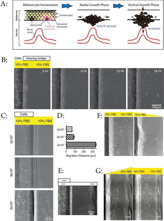
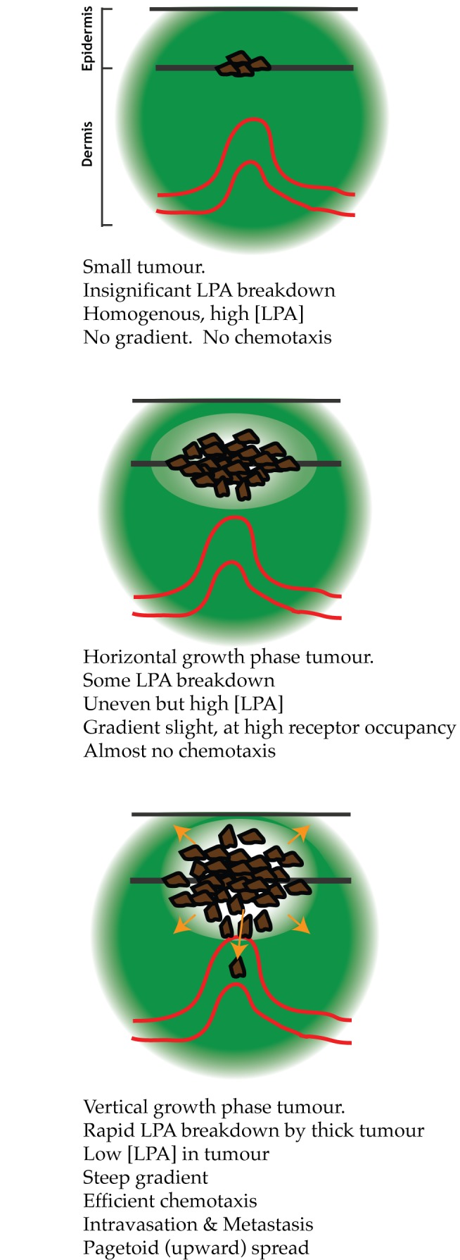
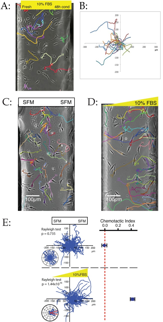
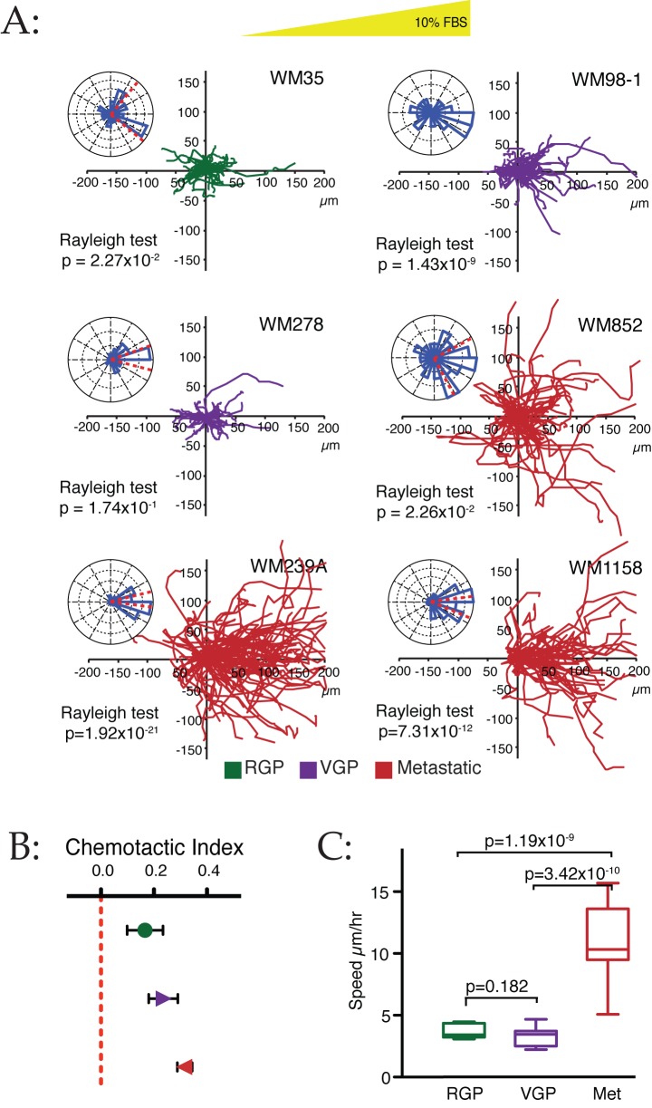
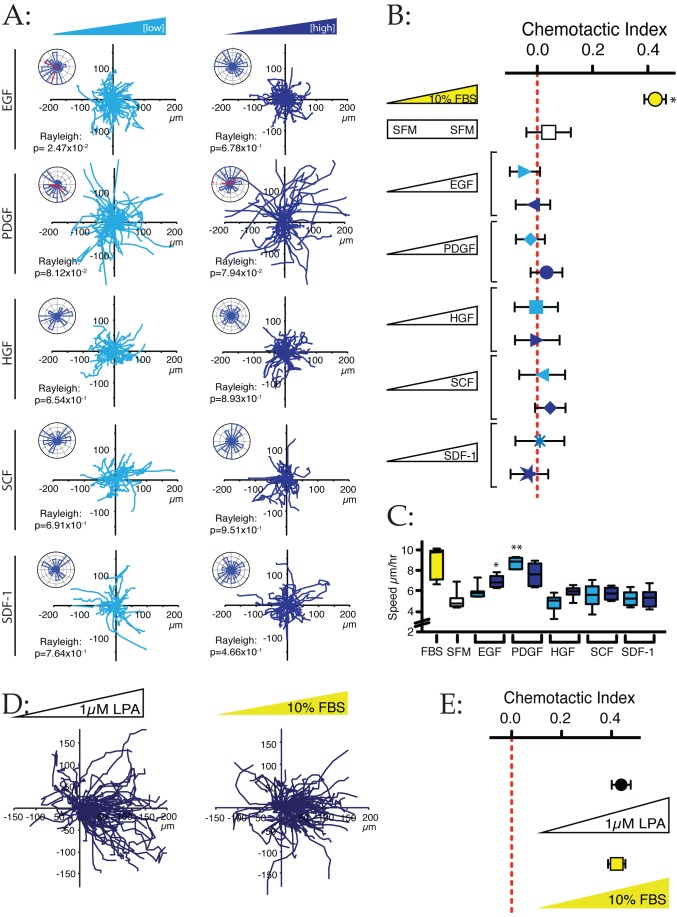
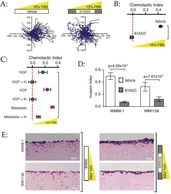
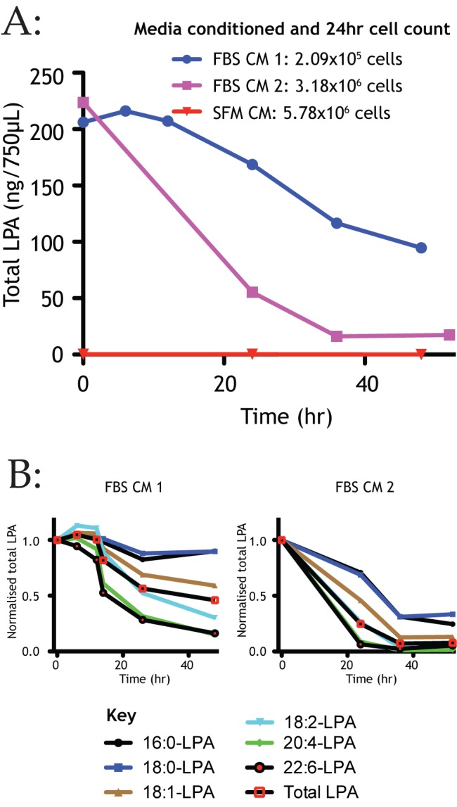
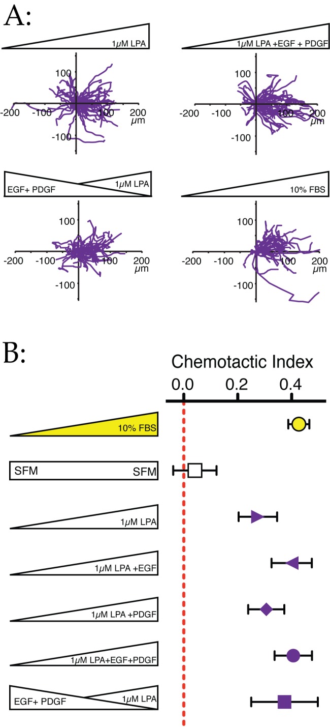
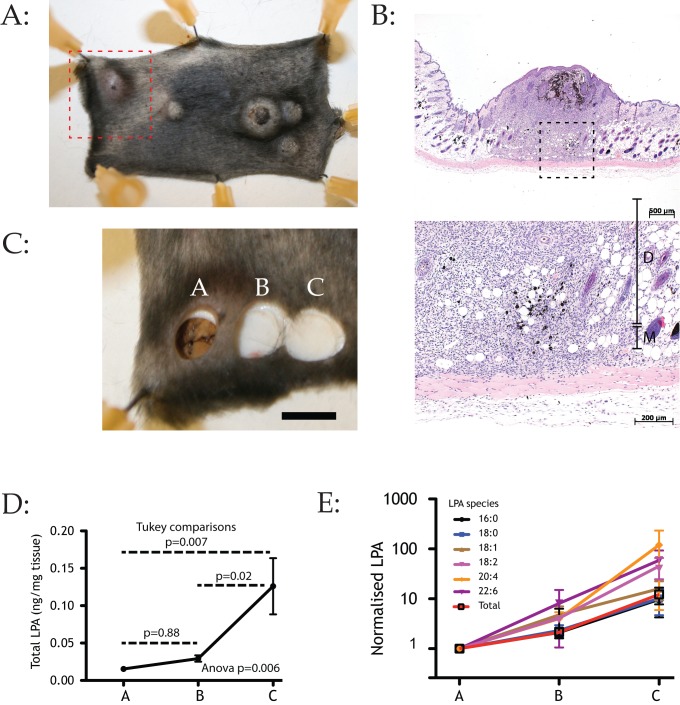
The high mortality of melanoma is caused by rapid spread of cancer cells, which occurs unusually early in tumour evolution. Unlike most solid tumours, thickness rather than cytological markers or differentiation is the best guide to metastatic potential. Multiple stimuli that drive melanoma cell migration have been described, but it is not clear which are responsible for invasion, nor if chemotactic gradients exist in real tumours. In a chamber-based assay for melanoma dispersal, we find that cells migrate efficiently away from one another, even in initially homogeneous medium. This dispersal is driven by positive chemotaxis rather than chemorepulsion or contact inhibition. The principal chemoattractant, unexpectedly active across all tumour stages, is the lipid agonist lysophosphatidic acid (LPA) acting through the LPA receptor LPAR1. LPA induces chemotaxis of remarkable accuracy, and is both necessary and sufficient for chemotaxis and invasion in 2-D and 3-D assays. Growth factors, often described as tumour attractants, cause negligible chemotaxis themselves, but potentiate chemotaxis to LPA. Cells rapidly break down LPA present at substantial levels in culture medium and normal skin to generate outward-facing gradients. We measure LPA gradients across the margins of melanomas in vivo, confirming the physiological importance of our results. We conclude that LPA chemotaxis provides a strong drive for melanoma cells to invade outwards. Cells create their own gradients by acting as a sink, breaking down locally present LPA, and thus forming a gradient that is low in the tumour and high in the surrounding areas. The key step is not acquisition of sensitivity to the chemoattractant, but rather the tumour growing to break down enough LPA to form a gradient. Thus the stimulus that drives cell dispersal is not the presence of LPA itself, but the self-generated, outward-directed gradient.
黑色素瘤的高死亡率是由癌细胞的快速扩散所致,这种扩散在肿瘤发展过程中异常早期就会发生。与大多数实体瘤不同,黑色素瘤转移潜能的最佳指标是肿瘤厚度,而非细胞学标志物或分化程度。虽然已经描述了多种驱动黑色素瘤细胞迁移的刺激因素,但尚不清楚哪些因素导致侵袭,也不清楚实体瘤中是否存在趋化梯度。在一项基于小室的黑色素瘤扩散试验中,我们发现细胞即使在最初均匀的培养基中也能有效地相互远离迁移。这种扩散是由正向趋化作用驱动的,而非化学排斥或接触抑制。出人意料的是,主要的趋化因子在所有肿瘤阶段均有活性,它是通过LPA受体LPAR1起作用的脂质激动剂溶血磷脂酸(LPA)。LPA诱导的趋化作用具有极高的准确性,在二维和三维试验中,它对于趋化作用和侵袭都是必需且充分的。通常被描述为肿瘤吸引剂的生长因子本身引起的趋化作用可忽略不计,但能增强对LPA的趋化作用。细胞会迅速分解培养基和正常皮肤中大量存在的LPA,以产生外向梯度。我们在体内测量了黑色素瘤边缘的LPA梯度,证实了我们结果的生理学重要性。我们得出结论,LPA趋化作用为黑色素瘤细胞向外侵袭提供了强大动力。细胞通过充当一个汇,分解局部存在的LPA,从而形成肿瘤内低而周围区域高的梯度,进而产生自身的梯度。关键步骤不是获得对趋化因子的敏感性,而是肿瘤生长到能够分解足够的LPA以形成梯度。因此,驱动细胞扩散的刺激因素不是LPA本身的存在,而是自我产生的外向梯度。