Department of Neurology, Bispebjerg Hospital, University of Copenhagen , Copenhagen , Denmark.
Department of Radiology, Bispebjerg Hospital, University of Copenhagen , Copenhagen , Denmark.
Front Neurol. 2014 Sep 29;5:186. doi: 10.3389/fneur.2014.00186. eCollection 2014.
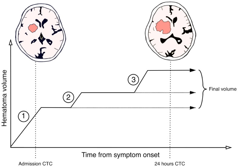
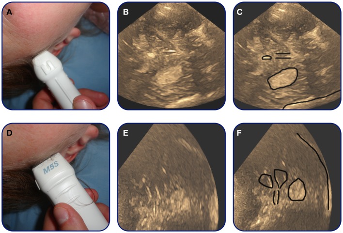
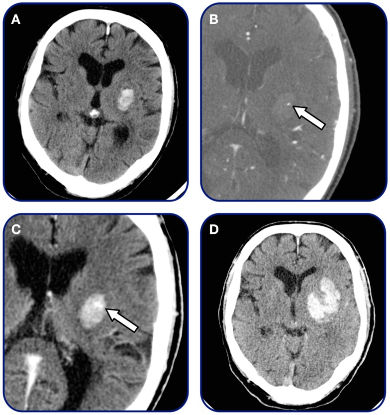
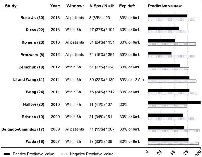
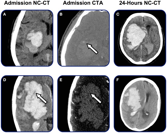
Post-admission hematoma expansion in patients with intracerebral hemorrhage (ICH) comprises a simultaneous major clinical problem and a possible target for medical intervention. In any case, the ability to predict and observe hematoma expansion is of great clinical importance. We review radiological concepts in predicting and observing post-admission hematoma expansion. Hematoma expansion can be observed within the first 24 h after symptom onset, but predominantly occurs in the early hours. Thus capturing markers of on-going bleeding on imaging techniques could predict hematoma expansion. The spot sign observed on computed tomography angiography is believed to represent on-going bleeding and is to date the most well investigated and reliable radiological predictor of hematoma expansion as well as functional outcome and mortality. On non-contrast CT, the presence of foci of hypoattenuation within the hematoma along with the hematoma-size is reported to be predictive of hematoma expansion and outcome. Because patients tend to arrive earlier to the hospital, a larger fraction of acute ICH-patients must be expected to undergo hematoma expansion. This renders observation and radiological follow-up investigations increasingly relevant. Transcranial duplex sonography has in recent years proven to be able to estimate hematoma volume with good precision and could be a valuable tool in bedside serial observation of acute ICH-patients. Future studies will elucidate, if better prediction and observation of post-admission hematoma expansion can help select patients, who will benefit from hemostatic treatment.
脑出血(ICH)患者的入院后血肿扩大是一个同时存在的重大临床问题,也是可能的医学干预目标。在任何情况下,预测和观察血肿扩大的能力都具有重要的临床意义。我们回顾了预测和观察入院后血肿扩大的影像学概念。血肿扩大可在症状出现后 24 小时内观察到,但主要发生在早期。因此,在影像学技术上捕捉到正在发生的出血的标志物可以预测血肿扩大。在 CT 血管造影上观察到的斑点征被认为代表正在发生的出血,是迄今为止研究最充分、最可靠的血肿扩大以及功能结局和死亡率的影像学预测指标。在非对比 CT 上,血肿内存在的低衰减灶与血肿大小一起被报道可预测血肿扩大和结局。由于患者往往更早到达医院,预计会有更多的急性 ICH 患者发生血肿扩大。这使得观察和影像学随访调查变得越来越重要。经颅双功超声检查近年来已被证明能够精确估计血肿量,可能成为急性 ICH 患者床边连续观察的有价值工具。未来的研究将阐明,对入院后血肿扩大的更好预测和观察是否有助于选择可能受益于止血治疗的患者。