Carmeli Cristian, Fornari Eleonora, Jalili Mahdi, Meuli Reto, Knyazeva Maria G
LREN, Department of Clinical Neuroscience, Centre Hospitalier Universitaire Vaudois (CHUV), University of Lausanne Lausanne, Switzerland.
Department of Radiology, Centre Hospitalier Universitaire Vaudois (CHUV), University of Lausanne Lausanne, Switzerland ; CIBM (Centre d'Imagérie Biomédicale), CHUV Unit Lausanne, Switzerland.
Brain Behav. 2014 Sep;4(5):721-37. doi: 10.1002/brb3.252. Epub 2014 Jul 28.
Interindividual variations in regional structural properties covary across the brain, thus forming networks that change as a result of aging and accompanying neurological conditions. The alterations of superficial white matter (SWM) in Alzheimer's disease (AD) are of special interest, since they follow the AD-specific pattern characterized by the strongest neurodegeneration of the medial temporal lobe and association cortices.
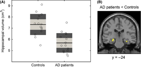
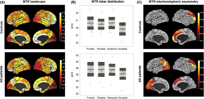
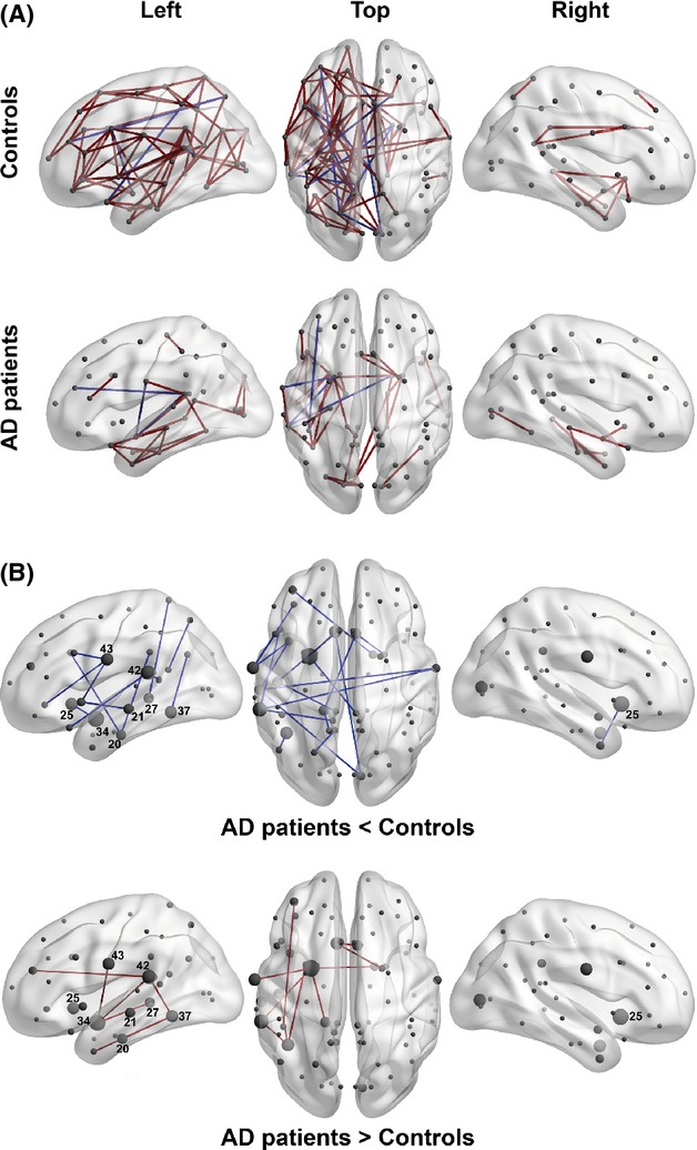
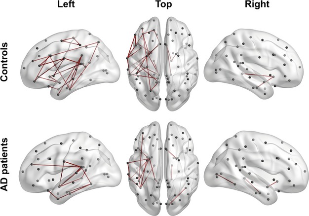
Here, we present an SWM network analysis in comparison with SWM topography based on the myelin content quantified with magnetization transfer ratio (MTR) for 39 areas in each hemisphere in 15 AD patients and 15 controls. The networks are represented by graphs, in which nodes correspond to the areas, and edges denote statistical associations between them.
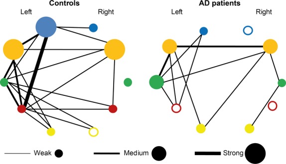
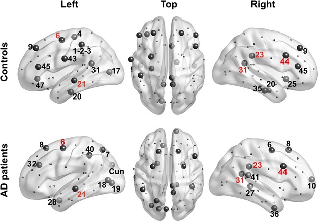
In both groups, the networks were characterized by asymmetrically distributed edges (predominantly in the left hemisphere). The AD-related differences were also leftward. The edges lost due to AD tended to connect nodes in the temporal lobe to other lobes or nodes within or between the latter lobes. The newly gained edges were mostly confined to the temporal and paralimbic regions, which manifest demyelination of SWM already in mild AD.
This pattern suggests that the AD pathological process coordinates SWM demyelination in the temporal and paralimbic regions, but not elsewhere. A comparison of the MTR maps with MTR-based networks shows that although, in general, the changes in network architecture in AD recapitulate the topography of (de)myelination, some aspects of structural covariance (including the interhemispheric asymmetry of networks) have no immediate reflection in the myelination pattern.
大脑区域结构特性的个体间差异相互关联,从而形成因衰老及伴随的神经疾病而发生变化的网络。阿尔茨海默病(AD)中脑表面白质(SWM)的改变尤其引人关注,因为它们遵循AD特有的模式,其特征是内侧颞叶和联合皮质的神经退行性变最为严重。
在此,我们呈现了一项SWM网络分析,并将其与基于髓鞘含量的SWM地形图进行比较,该髓鞘含量通过磁化传递率(MTR)对15名AD患者和15名对照者每个半球的39个区域进行量化。网络由图表示,其中节点对应于各个区域,边表示它们之间的统计关联。
在两组中,网络的特征均为边缘不对称分布(主要在左半球)。与AD相关的差异也偏向左侧。因AD而丢失的边往往将颞叶中的节点与其他叶或后叶内部或之间的节点相连。新获得的边大多局限于颞叶和边缘旁区域,这些区域在轻度AD中就已表现出SWM脱髓鞘。
这种模式表明,AD病理过程协调了颞叶和边缘旁区域的SWM脱髓鞘,但其他区域并非如此。将MTR图谱与基于MTR的网络进行比较表明,虽然一般来说,AD中网络结构的变化概括了(脱)髓鞘的地形图,但结构协方差的某些方面(包括网络的半球间不对称性)在髓鞘化模式中没有直接反映。