Department of Neurophysics, Max Planck Institute for Human Cognitive and Brain Sciences, 04103 Leipzig, Germany.
Department of Education and Psychology, Center for Cognitive Neuroscience Berlin, Free University Berlin, 14195 Berlin, Germany.
Cereb Cortex. 2020 Jun 30;30(8):4496-4514. doi: 10.1093/cercor/bhaa049.
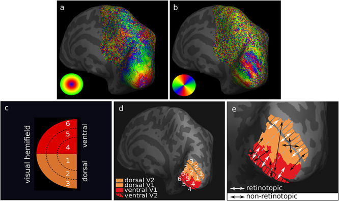
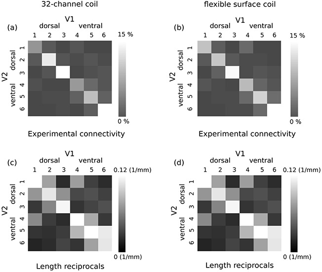
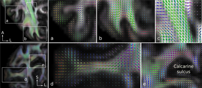
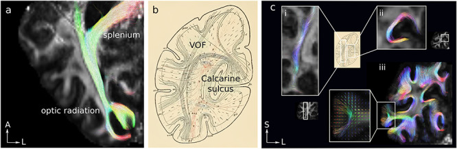
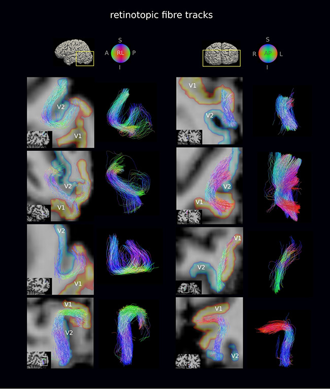
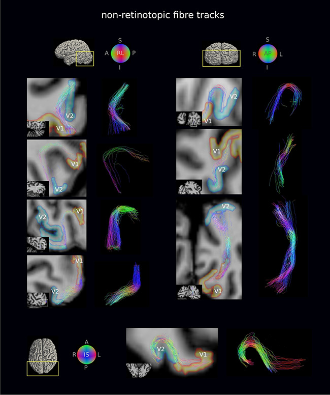
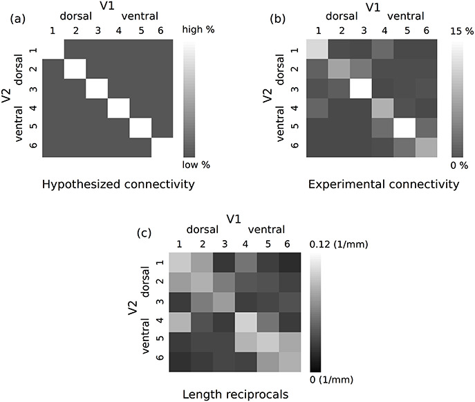
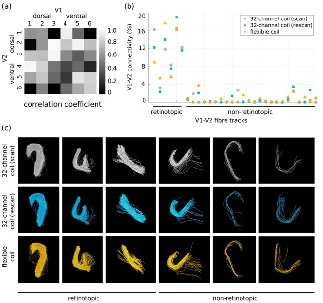
Short association fibers (U-fibers) connect proximal cortical areas and constitute the majority of white matter connections in the human brain. U-fibers play an important role in brain development, function, and pathology but are underrepresented in current descriptions of the human brain connectome, primarily due to methodological challenges in diffusion magnetic resonance imaging (dMRI) of these fibers. High spatial resolution and dedicated fiber and tractography models are required to reliably map the U-fibers. Moreover, limited quantitative knowledge of their geometry and distribution makes validation of U-fiber tractography challenging. Submillimeter resolution diffusion MRI-facilitated by a cutting-edge MRI scanner with 300 mT/m maximum gradient amplitude-was used to map U-fiber connectivity between primary and secondary visual cortical areas (V1 and V2, respectively) in vivo. V1 and V2 retinotopic maps were obtained using functional MRI at 7T. The mapped V1-V2 connectivity was retinotopically organized, demonstrating higher connectivity for retinotopically corresponding areas in V1 and V2 as expected. The results were highly reproducible, as demonstrated by repeated measurements in the same participants and by an independent replication group study. This study demonstrates a robust U-fiber connectivity mapping in vivo and is an important step toward construction of a more complete human brain connectome.
短连合纤维(U 纤维)连接近端皮质区,构成人类大脑白质连接的大部分。U 纤维在大脑发育、功能和病理学中发挥着重要作用,但在当前人类脑连接组的描述中代表性不足,主要是由于这些纤维的扩散磁共振成像(dMRI)方法存在挑战。需要高空间分辨率和专门的纤维和束追踪模型来可靠地绘制 U 纤维。此外,其几何形状和分布的定量知识有限,使得 U 纤维束追踪的验证具有挑战性。亚毫米分辨率的扩散 MRI 由具有 300 mT/m 最大梯度幅度的尖端 MRI 扫描仪辅助完成,用于在体内绘制初级和次级视觉皮质区(分别为 V1 和 V2)之间的 U 纤维连接。在 7T 下使用功能磁共振成像获得 V1 和 V2 的视网膜映射图。绘制的 V1-V2 连接具有视网膜组织特异性,与预期一样,V1 和 V2 中具有对应视网膜区域的连接更高。正如在同一参与者的重复测量和独立复制组研究中所证明的那样,结果具有高度可重复性。这项研究证明了在体内进行稳健的 U 纤维连接映射是构建更完整的人类脑连接组的重要一步。