Veale Thomas, Malone Ian B, Poole Teresa, Parker Thomas D, Slattery Catherine F, Paterson Ross W, Foulkes Alexander J M, Thomas David L, Schott Jonathan M, Zhang Hui, Fox Nick C, Cash David M
The Dementia Research Centre, Department of Neurodegenerative Disease, UCL Queen Square Institute of Neurology, London, UK.
UK Dementia Research Institute at UCL, University College London, London, UK.
Brain Commun. 2021 Nov 15;3(4):fcab272. doi: 10.1093/braincomms/fcab272. eCollection 2021.
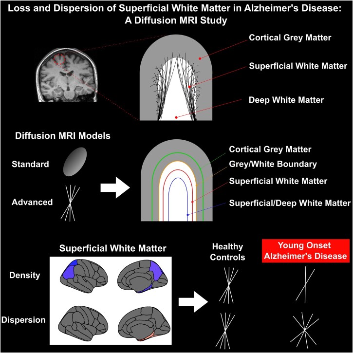
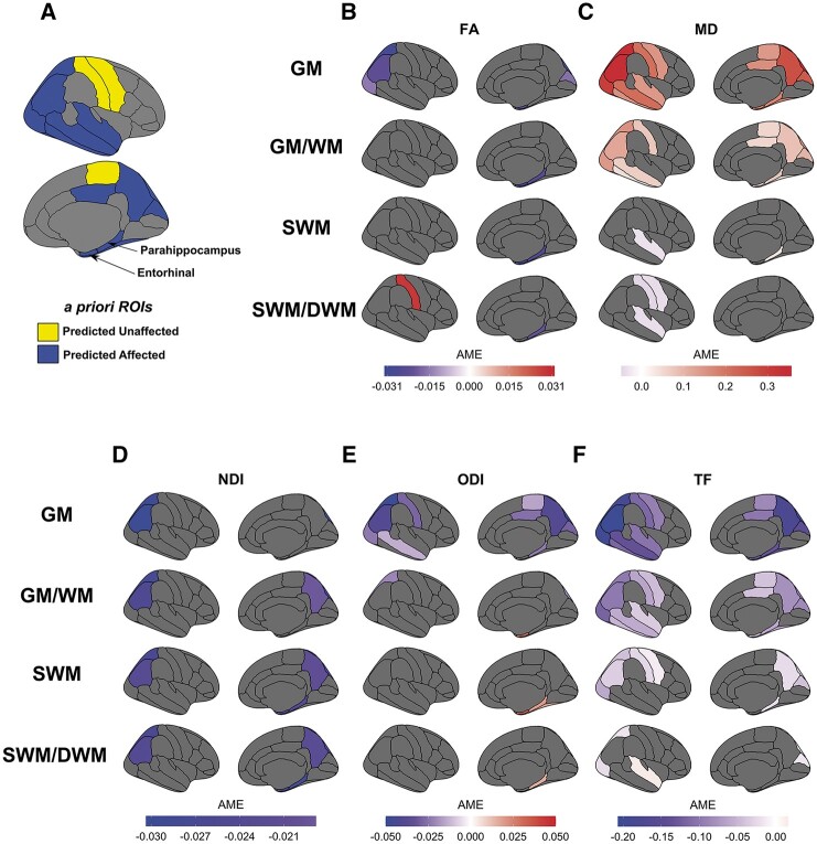
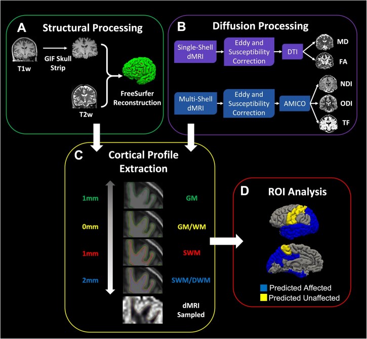
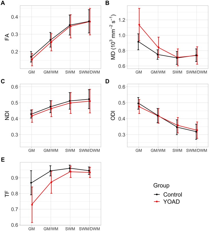
Pathological cerebral white matter changes in Alzheimer's disease have been shown using diffusion tensor imaging. Superficial white matter changes are relatively understudied despite their importance in cortico-cortical connections. Measuring superficial white matter degeneration using diffusion tensor imaging is challenging due to its complex organizational structure and proximity to the cortex. To overcome this, we investigated diffusion MRI changes in young-onset Alzheimer's disease using standard diffusion tensor imaging and Neurite Orientation Dispersion and Density Imaging to distinguish between disease-related changes that are degenerative (e.g. loss of myelinated fibres) and organizational (e.g. increased fibre dispersion). Twenty-nine young-onset Alzheimer's disease patients and 22 healthy controls had both single-shell and multi-shell diffusion MRI. We calculated fractional anisotropy, mean diffusivity, neurite density index, orientation dispersion index and tissue fraction (1-free water fraction). Diffusion metrics were sampled in 15 regions of interest at four points along the cortical profile: cortical grey matter, grey/white boundary, superficial white matter (1 mm below grey/white boundary) and superficial/deeper white matter (2 mm below grey/white boundary). To estimate cross-sectional group differences, we used average marginal effects from linear mixed effect models of participants' diffusion metrics along the cortical profile. The superficial white matter of young-onset Alzheimer's disease individuals had lower neurite density index compared to controls in five regions (superior and inferior parietal, precuneus, entorhinal and parahippocampus) (all <0.05), and higher orientation dispersion index in three regions (fusiform, entorhinal and parahippocampus) (all <0.05). Young-onset Alzheimer's disease individuals had lower fractional anisotropy in the entorhinal and parahippocampus regions (both <0.05) and higher fractional anisotropy within the postcentral region (<0.05). Mean diffusivity was higher in the young-onset Alzheimer's disease group in the parahippocampal region (<0.05) and lower in the postcentral, precentral and superior temporal regions (all <0.05). In the overlying grey matter, disease-related changes were largely consistent with superficial white matter findings when using neurite density index and fractional anisotropy, but appeared at odds with orientation dispersion and mean diffusivity. Tissue fraction was significantly lower across all grey matter regions in young-onset Alzheimer's disease individuals (all <0.001) but group differences reduced in magnitude and coverage when moving towards the superficial white matter. These results show that microstructural changes occur within superficial white matter and along the cortical profile in individuals with young-onset Alzheimer's disease. Lower neurite density and higher orientation dispersion suggests underlying fibres undergo neurodegeneration and organizational changes, two effects previously indiscernible using standard diffusion tensor metrics in superficial white matter.
使用扩散张量成像已显示出阿尔茨海默病中病理性脑白质变化。尽管浅层白质变化在皮质 - 皮质连接中很重要,但相对而言研究较少。由于其组织结构复杂且靠近皮质,使用扩散张量成像测量浅层白质变性具有挑战性。为了克服这一问题,我们使用标准扩散张量成像和神经突方向离散度与密度成像研究了早发型阿尔茨海默病中的扩散磁共振成像变化,以区分与疾病相关的退行性变化(如髓鞘纤维丢失)和组织学变化(如纤维离散度增加)。29例早发型阿尔茨海默病患者和22名健康对照者均进行了单壳和多壳扩散磁共振成像。我们计算了分数各向异性、平均扩散率、神经突密度指数、方向离散度指数和组织分数(1 - 自由水分数)。在沿皮质轮廓的四个点的15个感兴趣区域中采样扩散指标:皮质灰质、灰/白质边界、浅层白质(灰/白质边界下方1毫米)和浅层/深层白质(灰/白质边界下方2毫米)。为了估计组间横断面差异,我们使用了参与者沿皮质轮廓的扩散指标的线性混合效应模型的平均边际效应。早发型阿尔茨海默病个体的浅层白质在五个区域(顶叶上下部、楔前叶、内嗅区和海马旁回)的神经突密度指数低于对照组(均<0.05),在三个区域(梭状回、内嗅区和海马旁回)的方向离散度指数较高(均<0.05)。早发型阿尔茨海默病个体在内嗅区和海马旁回区域的分数各向异性较低(均<0.05),而中央后回区域内的分数各向异性较高(<0.05)。早发型阿尔茨海默病组海马旁回区域的平均扩散率较高(<0.05),而中央后回、中央前回和颞上回区域的平均扩散率较低(均<0.05)。在覆盖的灰质中,当使用神经突密度指数和分数各向异性时,与疾病相关的变化在很大程度上与浅层白质的发现一致,但与方向离散度和平均扩散率似乎不一致。早发型阿尔茨海默病个体所有灰质区域的组织分数均显著较低(均<0.001),但当向浅层白质移动时,组间差异的幅度和范围减小。这些结果表明,早发型阿尔茨海默病个体的浅层白质内以及沿皮质轮廓发生了微观结构变化。较低的神经突密度和较高的方向离散度表明潜在纤维发生了神经退行性变和组织学变化,这两种效应以前使用标准扩散张量指标在浅层白质中难以区分。