Le Phong T, Pearce Meghan M, Zhang Shubin, Campbell Edward M, Fok Cynthia S, Mueller Elizabeth R, Brincat Cynthia A, Wolfe Alan J, Brubaker Linda
Department of Microbiology and Immunology, Stritch School of Medicine, Loyola University Chicago, Maywood, Illinois, United States of America.
University of Minnesota, Department of Urology, Minneapolis, Minnesota, United States of America.
PLoS One. 2014 Oct 29;9(10):e111375. doi: 10.1371/journal.pone.0111375. eCollection 2014.
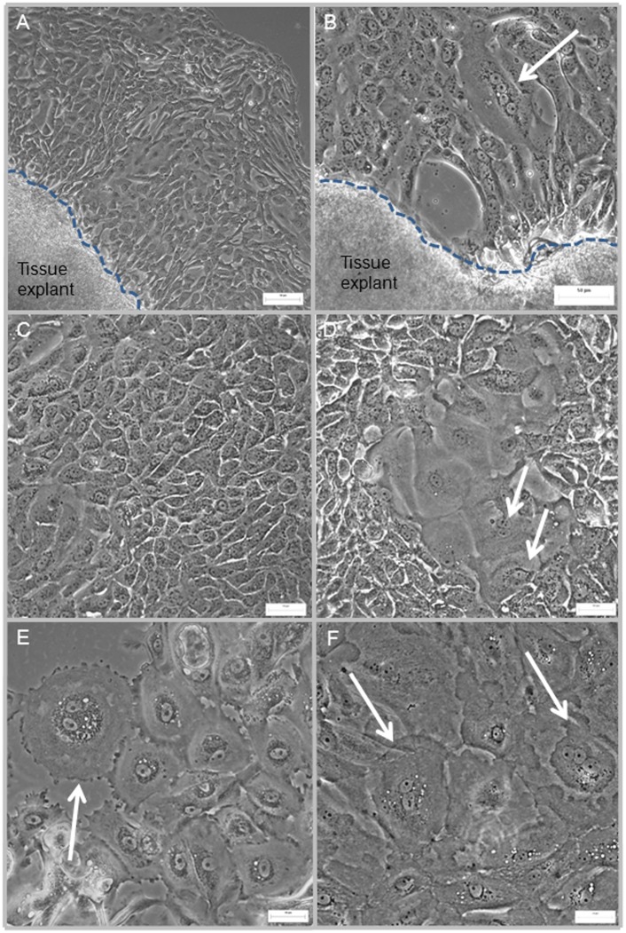
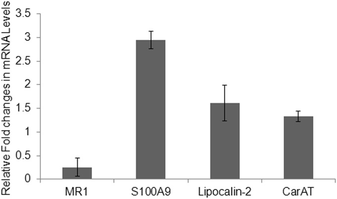
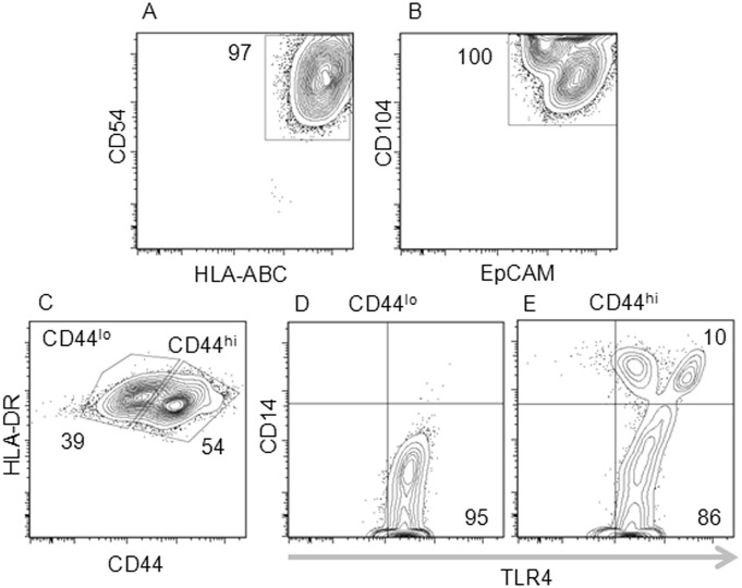
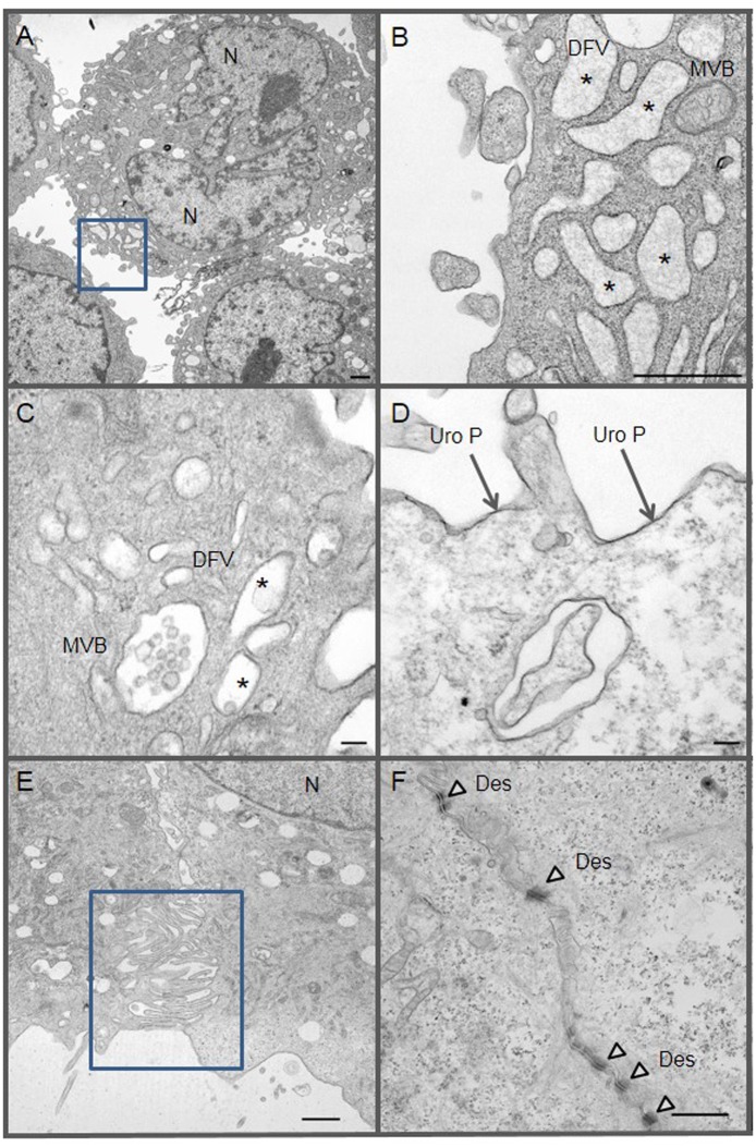
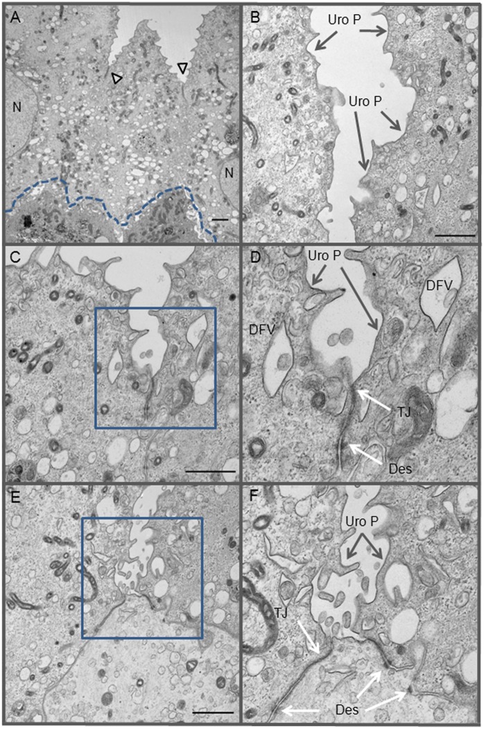
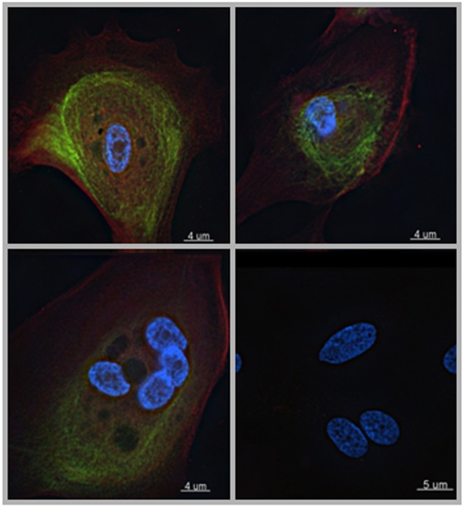
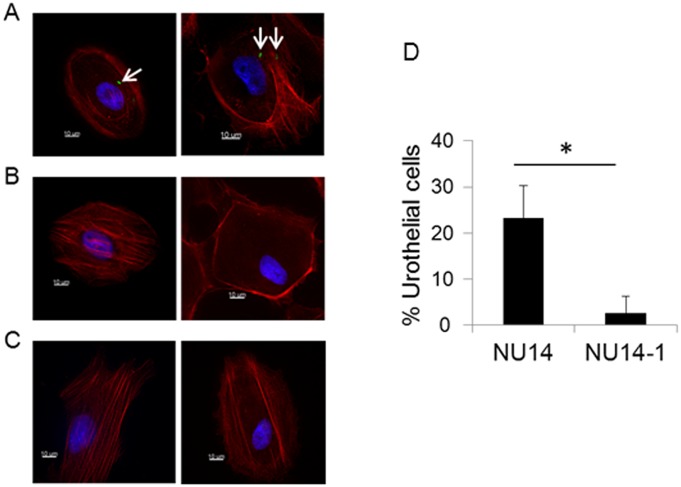
Human urinary disorders are generally studied in rodent models due to limitations of functional in vitro culture models of primary human urothelial cells (HUCs). Current HUC culture models are often derived from immortalized cancer cell lines, which likely have functional characteristics differ from healthy human urothelium. Here, we described a simple explant culture technique to generate HUCs and assessed their in vitro functions. Using transmission electron microscopy, we assessed morphology and heterogeneity of the generated HUCs and characterized their intercellular membrane structural proteins relative to ex vivo urothelium tissue. We demonstrated that our cultured HUCs are free of fibroblasts. They are also heterogeneous, containing cells characteristic of both immature basal cells and mature superficial urothelial cells. The cultured HUCs expressed muscarinic receptors (MR1 and MR2), carnitine acetyltransferase (CarAT), immunoregulatory cytokines IL7, IL15, and IL23, as well as the chemokine CCL20. HUCs also expressed epithelial cell-specific molecules essential for forming intercellular structures that maintain the functional capacity to form the physiological barrier of the human bladder urothelium. A subset of HUCs, identified by the high expression of CD44, expressed the Toll-like receptor 4 (TLR4) along with its co-receptor CD14. We demonstrated that HUCs express, at the mRNA level, both forms of the IL22 receptor, the membrane-associated (IL22RA1) and the secreted soluble (IL22RA2) forms; in turn, IL22 inhibited expression of MR1 and induced expression of CarAT and two antimicrobial peptides (S100A9 and lipocalin-2). While the cellular sources of IL22 have yet to be identified, the HUC cytokine and chemokine profiles support the concept that IL22-producing cells are present in the human bladder mucosa tissue and that IL22 plays a regulatory role in HUC functions. Thus, the described explant technique is clearly capable of generating functional HUCs suitable for the study of human urinary tract disorders, including interactions between urothelium and IL22-producing cells.
由于原代人尿路上皮细胞(HUCs)的功能性体外培养模型存在局限性,人类泌尿系统疾病通常在啮齿动物模型中进行研究。当前的HUC培养模型通常来源于永生化癌细胞系,其功能特性可能与健康的人尿路上皮不同。在此,我们描述了一种简单的外植体培养技术来生成HUCs,并评估了它们的体外功能。使用透射电子显微镜,我们评估了所生成HUCs的形态和异质性,并相对于离体尿路上皮组织对其细胞间膜结构蛋白进行了表征。我们证明我们培养的HUCs不含成纤维细胞。它们也是异质性的,包含未成熟基底细胞和成熟表层尿路上皮细胞的特征性细胞。培养的HUCs表达毒蕈碱受体(MR1和MR2)、肉碱乙酰转移酶(CarAT)、免疫调节细胞因子IL7、IL15和IL23,以及趋化因子CCL20。HUCs还表达了形成细胞间结构所必需的上皮细胞特异性分子,这些结构维持了形成人膀胱尿路上皮生理屏障的功能能力。通过CD44高表达鉴定的一部分HUCs表达了Toll样受体4(TLR4)及其共受体CD14。我们证明HUCs在mRNA水平表达两种形式的IL22受体,即膜相关形式(IL22RA1)和分泌的可溶性形式(IL22RA2);反过来,IL22抑制MR1的表达并诱导CarAT和两种抗菌肽(S100A9和lipocalin-2)的表达。虽然IL22的细胞来源尚未确定,但HUC细胞因子和趋化因子谱支持这样的概念,即产生IL22的细胞存在于人膀胱黏膜组织中,并且IL22在HUC功能中起调节作用。因此,所描述的外植体技术显然能够生成适用于研究人类泌尿系统疾病的功能性HUCs,包括尿路上皮与产生IL22的细胞之间的相互作用。