Medhora Meetha, Gao Feng, Glisch Chad, Narayanan Jayashree, Sharma Ashish, Harmann Leanne M, Lawlor Michael W, Snyder Laura A, Fish Brian L, Down Julian D, Moulder John E, Strande Jennifer L, Jacobs Elizabeth R
Department of Radiation Oncology, Medical College of Wisconsin, 8701, Watertown Plank Road, Milwaukee, WI 53226, USA Cardiovascular Center, Medical College of Wisconsin, 8701, Watertown Plank Road, Milwaukee, WI 53226, USA Division of Pulmonary Medicine, Medical College of Wisconsin, 8701, Watertown Plank Road, Milwaukee, WI 53226, USA Research Service, Department of Veteran's Affairs, Clement J. Zablocki VA Medical Center, Milwaukee, Wisconsin, USA
Department of Radiation Oncology, Medical College of Wisconsin, 8701, Watertown Plank Road, Milwaukee, WI 53226, USA.
J Radiat Res. 2015 Mar;56(2):248-60. doi: 10.1093/jrr/rru095. Epub 2014 Nov 3.
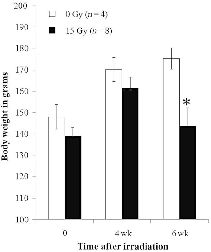
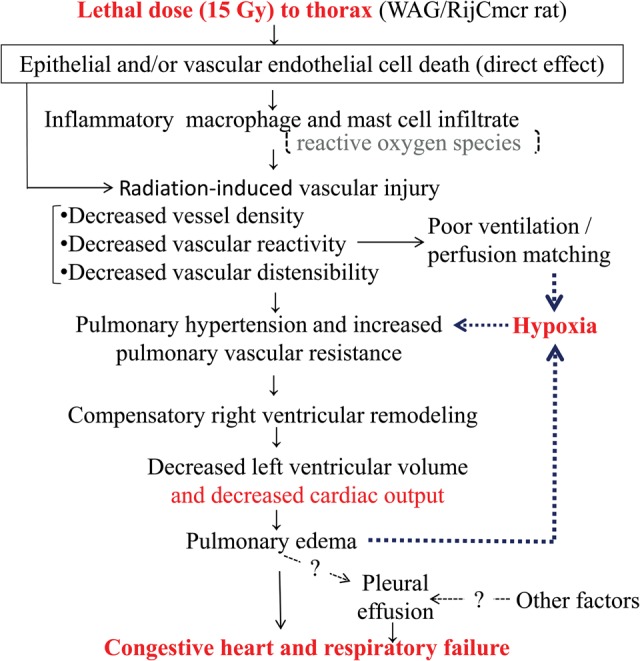
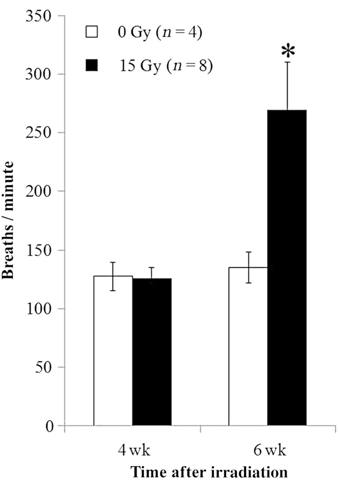
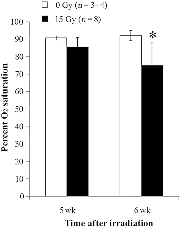
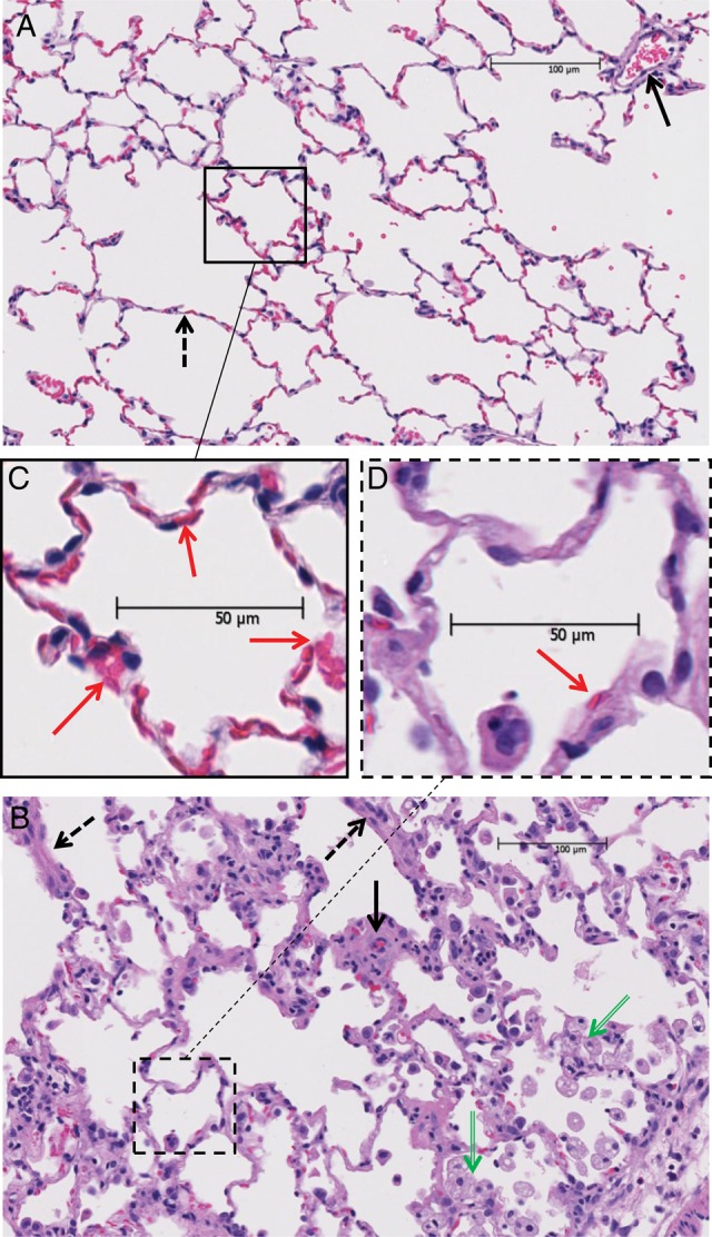
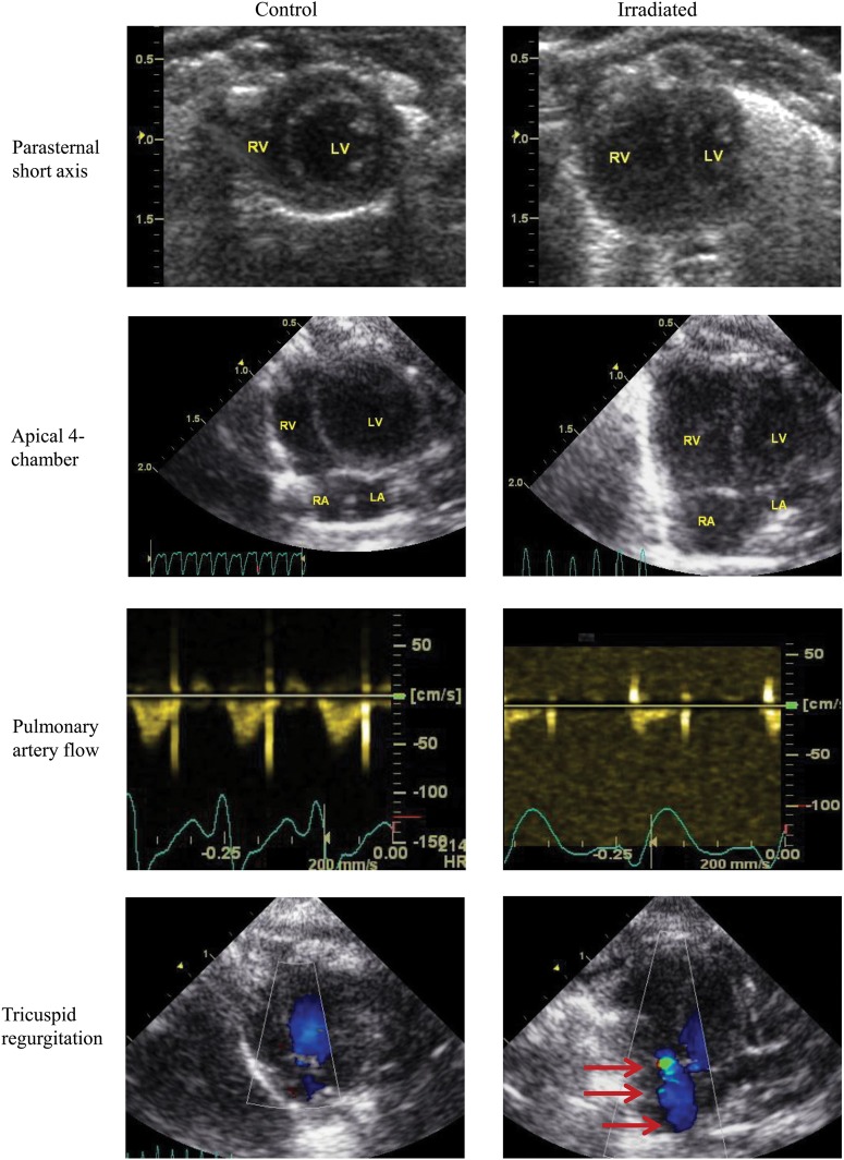
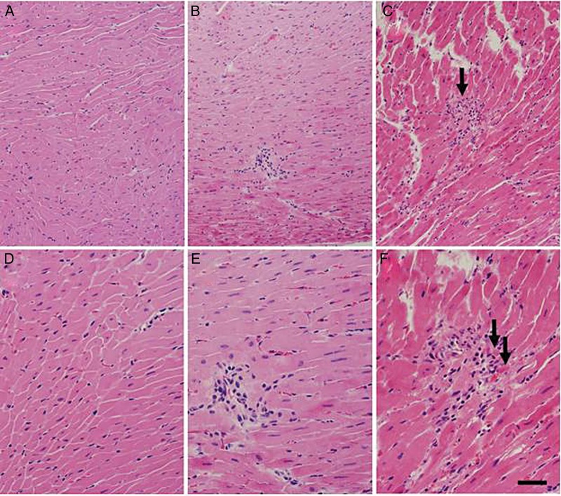
To study the mechanisms of death following a single lethal dose of thoracic radiation, WAG/RijCmcr (Wistar) rats were treated with 15 Gy to the whole thorax and followed until they were morbid or sacrificed for invasive assays at 6 weeks. Lung function was assessed by breathing rate and arterial oxygen saturation. Lung structure was evaluated histologically. Cardiac structure and function were examined by echocardiography. The frequency and characteristics of pleural effusions were determined. Morbidity from 15 Gy radiation occurred in all rats 5 to 8 weeks after exposure, coincident with histological pneumonitis. Increases in breathing frequencies peaked at 6 weeks, when profound arterial hypoxia was also recorded. Echocardiography analysis at 6 weeks showed pulmonary hypertension and severe right ventricular enlargement with impaired left ventricular function and cardiac output. Histologic sections of the heart revealed only rare foci of lymphocytic infiltration. Total lung weight more than doubled. Pleural effusions were present in the majority of the irradiated rats and contained elevated protein, but low lactate dehydrogenase, when compared with serum from the same animal. Pleural effusions had a higher percentage of macrophages and large monocytes than neutrophils and contained mast cells that are rarely present in other pathological states. Lethal irradiation to rat lungs leads to hypoxia with infiltration of immune cells, edema and pleural effusion. These changes may contribute to pulmonary vascular and parenchymal injury that result in secondary changes in heart structure and function. We report that conditions resembling congestive heart failure contribute to death during radiation pneumonitis, which indicates new targets for therapy.
为研究单次致死剂量胸部放疗后的死亡机制,对WAG/RijCmcr(Wistar)大鼠进行全胸15 Gy照射,并随访至6周时出现病态或为进行侵入性检测而处死。通过呼吸频率和动脉血氧饱和度评估肺功能。组织学评估肺结构。通过超声心动图检查心脏结构和功能。确定胸腔积液的频率和特征。15 Gy辐射导致所有大鼠在照射后5至8周发病,同时出现组织学肺炎。呼吸频率增加在6周时达到峰值,此时也记录到严重的动脉低氧血症。6周时的超声心动图分析显示肺动脉高压和严重的右心室扩大,左心室功能和心输出量受损。心脏组织学切片仅显示罕见的淋巴细胞浸润灶。肺总重量增加了一倍多。大多数受照射大鼠出现胸腔积液,与同一动物的血清相比,胸腔积液中蛋白质含量升高,但乳酸脱氢酶含量低。胸腔积液中巨噬细胞和大单核细胞的比例高于中性粒细胞,且含有在其他病理状态下很少出现的肥大细胞。对大鼠肺部进行致死性照射会导致低氧血症,并伴有免疫细胞浸润、水肿和胸腔积液。这些变化可能导致肺血管和实质损伤,进而引起心脏结构和功能的继发性改变。我们报告,类似充血性心力衰竭的情况会导致放射性肺炎期间的死亡,这为治疗指明了新的靶点。