• 文献检索
  • 文档翻译
  • 深度研究
  • 学术资讯
  • Suppr Zotero 插件Zotero 插件
  • 邀请有礼
  • 套餐&价格
  • 历史记录
应用&插件
Suppr Zotero 插件Zotero 插件浏览器插件Mac 客户端Windows 客户端微信小程序
定价
高级版会员购买积分包购买API积分包
服务
文献检索文档翻译深度研究API 文档MCP 服务
关于我们
关于 Suppr公司介绍联系我们用户协议隐私条款
关注我们

Suppr 超能文献

核心技术专利:CN118964589B侵权必究
粤ICP备2023148730 号-1Suppr @ 2026

文献检索

告别复杂PubMed语法,用中文像聊天一样搜索,搜遍4000万医学文献。AI智能推荐,让科研检索更轻松。

立即免费搜索

文件翻译

保留排版,准确专业,支持PDF/Word/PPT等文件格式,支持 12+语言互译。

免费翻译文档

深度研究

AI帮你快速写综述,25分钟生成高质量综述,智能提取关键信息,辅助科研写作。

立即免费体验

利用微阵列分析研究胰岛素对破骨细胞前体细胞基因表达谱的影响。

Gene expression profiling in osteoclast precursors by insulin using microarray analysis.

作者信息

Kim Hong Sung, Lee Na Kyung

机构信息

Department of Biomedical Laboratory Science, College of Medical Sciences, Soonchunhyang University, Asan 336-745, Korea.

出版信息

Mol Cells. 2014 Nov;37(11):827-32. doi: 10.14348/molcells.2014.0223. Epub 2014 Nov 5.

DOI:10.14348/molcells.2014.0223
PMID:25377254
原文链接:https://pmc.ncbi.nlm.nih.gov/articles/PMC4255103/
Abstract

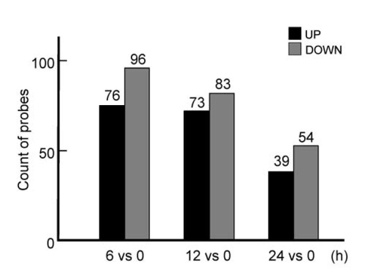
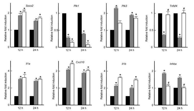
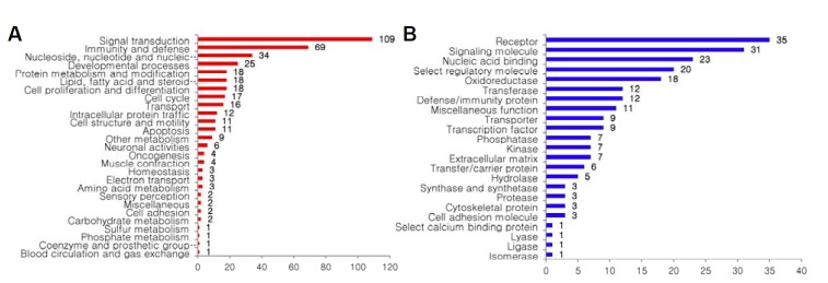
The balance between bone formation by osteoblasts and destruction of mineralized bone matrix by osteoclasts is important for bone homeostasis. The increase of osteoclast differentiation by RANKL induces bone diseases such as osteoporosis. Recent studies have shown that insulin is one of main factors mediating the cross-talk between bone remodeling and energy metabolism. However, the systemic examination of insulin-induced differential gene expression profiles in osteoclasts has not been extensively studied. Here, we investigated the global effects of insulin on osteoclast precursors at the level of gene transcription by microarray analysis. The number of genes that were up-regulated by ≥ 1.5 fold after insulin treatment for 6 h, 12 h, or 24 h was 76, 73, and 39; and 96, 83, and 54 genes were down-regulated, respectively. The genes were classified by 20 biological processes or 24 molecular functions and the number of genes involved in 'development processes' and 'cell proliferation and differentiation' was 25 and 18, respectively, including Inhba, Socs, Plk3, Tnfsf4, and Plk1. The microarray results of these genes were verified by real-time RT-PCR analysis. We also compared the effects of insulin and RANKL on the expression of these genes. Most genes had a very similar pattern of expressions in insulin- and RANKL-treated cells. Interestingly, Tnfsf4 and Inhba genes were affected by insulin but not by RANKL. Taken together, these results suggest a potential role for insulin in osteoclast biology, thus contributing to the understanding of the pathogenesis and development of therapeutics for numerous bone and metabolic diseases.

摘要

成骨细胞形成骨与破骨细胞破坏矿化骨基质之间的平衡对于骨稳态至关重要。RANKL诱导破骨细胞分化增加会引发骨质疏松等骨疾病。最近的研究表明,胰岛素是介导骨重塑与能量代谢之间相互作用的主要因素之一。然而,胰岛素诱导破骨细胞中差异基因表达谱的系统研究尚未广泛开展。在此,我们通过微阵列分析在基因转录水平上研究了胰岛素对破骨细胞前体的整体影响。胰岛素处理6小时、12小时或24小时后上调≥1.5倍的基因数量分别为76个、73个和39个;下调的基因数量分别为96个、83个和54个。这些基因按20个生物学过程或24个分子功能进行分类,参与“发育过程”和“细胞增殖与分化”的基因数量分别为25个和18个,包括Inhba、Socs、Plk3、Tnfsf4和Plk1。这些基因的微阵列结果通过实时RT-PCR分析进行了验证。我们还比较了胰岛素和RANKL对这些基因表达的影响。大多数基因在胰岛素和RANKL处理的细胞中具有非常相似的表达模式。有趣的是,Tnfsf4和Inhba基因受胰岛素影响,但不受RANKL影响。综上所述,这些结果表明胰岛素在破骨细胞生物学中具有潜在作用,从而有助于理解多种骨和代谢疾病的发病机制及治疗方法的开发。

https://cdn.ncbi.nlm.nih.gov/pmc/blobs/c629/4255103/f736a364091b/molcell-37-11-827f3.jpg
https://cdn.ncbi.nlm.nih.gov/pmc/blobs/c629/4255103/2e38748a7426/molcell-37-11-827f1.jpg
https://cdn.ncbi.nlm.nih.gov/pmc/blobs/c629/4255103/e8124a065ce1/molcell-37-11-827f2.jpg
https://cdn.ncbi.nlm.nih.gov/pmc/blobs/c629/4255103/f736a364091b/molcell-37-11-827f3.jpg
https://cdn.ncbi.nlm.nih.gov/pmc/blobs/c629/4255103/2e38748a7426/molcell-37-11-827f1.jpg
https://cdn.ncbi.nlm.nih.gov/pmc/blobs/c629/4255103/e8124a065ce1/molcell-37-11-827f2.jpg
https://cdn.ncbi.nlm.nih.gov/pmc/blobs/c629/4255103/f736a364091b/molcell-37-11-827f3.jpg

相似文献

1
Gene expression profiling in osteoclast precursors by insulin using microarray analysis.利用微阵列分析研究胰岛素对破骨细胞前体细胞基因表达谱的影响。
Mol Cells. 2014 Nov;37(11):827-32. doi: 10.14348/molcells.2014.0223. Epub 2014 Nov 5.
2
Aging increases stromal/osteoblastic cell-induced osteoclastogenesis and alters the osteoclast precursor pool in the mouse.衰老会增加基质/成骨细胞诱导的破骨细胞生成,并改变小鼠体内破骨细胞前体细胞库。
J Bone Miner Res. 2005 Sep;20(9):1659-68. doi: 10.1359/JBMR.050503. Epub 2005 May 2.
3
Expression profiling of microRNAs in RAW264.7 cells treated with a combination of tumor necrosis factor alpha and RANKL during osteoclast differentiation.在破骨细胞分化过程中,用肿瘤坏死因子 α 和 RANKL 联合处理 RAW264.7 细胞,对 microRNAs 进行表达谱分析。
J Periodontal Res. 2013 Jun;48(3):373-85. doi: 10.1111/jre.12017. Epub 2012 Oct 18.
4
A medium-chain fatty acid, capric acid, inhibits RANKL-induced osteoclast differentiation via the suppression of NF-κB signaling and blocks cytoskeletal organization and survival in mature osteoclasts.一种中链脂肪酸,癸酸,通过抑制核因子κB信号通路来抑制核因子κB受体活化因子配体(RANKL)诱导的破骨细胞分化,并阻断成熟破骨细胞的细胞骨架组织和存活。
Mol Cells. 2014 Aug;37(8):598-604. doi: 10.14348/molcells.2014.0153. Epub 2014 Aug 18.
5
Coptisine inhibits RANKL-induced NF-κB phosphorylation in osteoclast precursors and suppresses function through the regulation of RANKL and OPG gene expression in osteoblastic cells.小檗碱通过调节成骨细胞中 RANKL 和 OPG 基因的表达,抑制破骨细胞前体中 RANKL 诱导的 NF-κB 磷酸化,从而抑制其功能。
J Nat Med. 2012 Jan;66(1):8-16. doi: 10.1007/s11418-011-0537-7. Epub 2011 Jun 9.
6
Arecoline suppresses RANKL-induced osteoclast differentiation in vitro and attenuates LPS-induced bone loss in vivo.槟榔碱可抑制体外 RANKL 诱导的破骨细胞分化,并减轻体内 LPS 诱导的骨丢失。
Phytomedicine. 2020 Apr;69:153195. doi: 10.1016/j.phymed.2020.153195. Epub 2020 Feb 22.
7
The collection of NFATc1-dependent transcripts in the osteoclast includes numerous genes non-essential to physiologic bone resorption.破骨细胞中 NFATc1 依赖性转录本的集合包括许多对生理骨吸收非必需的基因。
Bone. 2012 Nov;51(5):902-12. doi: 10.1016/j.bone.2012.08.113. Epub 2012 Aug 16.
8
Dual Role of Cyanidin-3-glucoside on the Differentiation of Bone Cells.矢车菊素-3-葡萄糖苷在骨细胞分化中的双重作用
J Dent Res. 2015 Dec;94(12):1676-83. doi: 10.1177/0022034515604620. Epub 2015 Sep 8.
9
[RANKL signaling and bone diseases. Quiescent osteoclast precursors and RANKL signaling].[核因子κB受体活化因子配体信号传导与骨疾病。静止破骨细胞前体与核因子κB受体活化因子配体信号传导]
Clin Calcium. 2011 Aug;21(8):1187-92.
10
Expression profile of RhoGTPases and RhoGEFs during RANKL-stimulated osteoclastogenesis: identification of essential genes in osteoclasts.RANKL刺激破骨细胞生成过程中RhoGTPases和RhoGEFs的表达谱:破骨细胞中关键基因的鉴定
J Bone Miner Res. 2006 Sep;21(9):1387-98. doi: 10.1359/jbmr.060613.

引用本文的文献

1
Protective effects of miR-24-2-5p in early stages of breast cancer bone metastasis.miR-24-2-5p在乳腺癌骨转移早期的保护作用。
Breast Cancer Res. 2024 Dec 18;26(1):186. doi: 10.1186/s13058-024-01934-2.
2
Alzheimer's Disease and Impaired Bone Microarchitecture, Regeneration and Potential Genetic Links.阿尔茨海默病与骨微结构受损、再生及潜在的遗传联系。
Life (Basel). 2023 Jan 29;13(2):373. doi: 10.3390/life13020373.
3
Trans-omics analysis of insulin action reveals a cell growth subnetwork which co-regulates anabolic processes.

本文引用的文献

1
Up-regulation of cyclinD1 and Bcl2A1 by insulin is involved in osteoclast proliferation.胰岛素对细胞周期蛋白D1和Bcl2A1的上调参与破骨细胞增殖。
Life Sci. 2014 Oct 2;114(2):57-61. doi: 10.1016/j.lfs.2014.07.006. Epub 2014 Jul 24.
2
OX40L blockade is therapeutic in arthritis, despite promoting osteoclastogenesis.OX40L 阻断在关节炎中具有治疗作用,尽管它促进了破骨细胞的生成。
Proc Natl Acad Sci U S A. 2014 Feb 11;111(6):2289-94. doi: 10.1073/pnas.1321071111. Epub 2014 Jan 27.
3
CD109 plays a role in osteoclastogenesis.CD109 在破骨细胞生成中发挥作用。
胰岛素作用的跨组学分析揭示了一个共同调节合成代谢过程的细胞生长子网。
iScience. 2022 Apr 8;25(5):104231. doi: 10.1016/j.isci.2022.104231. eCollection 2022 May 20.
4
Comprehensive Transcriptomic Profiling of Murine Osteoclast Differentiation Reveals Novel Differentially Expressed Genes and LncRNAs.小鼠破骨细胞分化的综合转录组分析揭示了新的差异表达基因和长链非编码RNA
Front Genet. 2021 Nov 15;12:781272. doi: 10.3389/fgene.2021.781272. eCollection 2021.
5
Integration of TGF-β-induced Smad signaling in the insulin-induced transcriptional response in endothelial cells.TGF-β 诱导的 Smad 信号在内皮细胞胰岛素诱导的转录反应中的整合。
Sci Rep. 2019 Nov 18;9(1):16992. doi: 10.1038/s41598-019-53490-x.
6
Trans-omic Analysis Reveals Selective Responses to Induced and Basal Insulin across Signaling, Transcriptional, and Metabolic Networks.跨组学分析揭示了信号转导、转录和代谢网络对诱导性和基础性胰岛素的选择性反应。
iScience. 2018 Sep 28;7:212-229. doi: 10.1016/j.isci.2018.07.022. Epub 2018 Sep 10.
7
Up-Regulation of RANK Expression via ERK1/2 by Insulin Contributes to the Enhancement of Osteoclast Differentiation.胰岛素通过ERK1/2上调RANK表达有助于增强破骨细胞分化。
Mol Cells. 2017 May 31;40(5):371-377. doi: 10.14348/molcells.2017.0025. Epub 2017 May 22.
8
Obatoclax regulates the proliferation and fusion of osteoclast precursors through the inhibition of ERK activation by RANKL.奥巴托克斯通过抑制RANKL激活ERK来调节破骨细胞前体的增殖和融合。
Mol Cells. 2015 Mar;38(3):279-84. doi: 10.14348/molcells.2015.2340. Epub 2015 Feb 4.
PLoS One. 2013 Apr 12;8(4):e61213. doi: 10.1371/journal.pone.0061213. Print 2013.
4
Caffeine enhances osteoclast differentiation and maturation through p38 MAP kinase/Mitf and DC-STAMP/CtsK and TRAP pathway.咖啡因通过 p38 MAP 激酶/Mitf 和 DC-STAMP/CtsK 及 TRAP 通路增强破骨细胞分化和成熟。
Cell Signal. 2013 May;25(5):1222-7. doi: 10.1016/j.cellsig.2013.02.015. Epub 2013 Feb 19.
5
Insulin stimulates gene expression of ferritin light chain in osteoblast cells.胰岛素刺激成骨细胞中铁蛋白轻链的基因表达。
J Cell Biochem. 2010 Dec 15;111(6):1493-500. doi: 10.1002/jcb.22879.
6
Insulin receptor signaling in osteoblasts regulates postnatal bone acquisition and body composition.成骨细胞中的胰岛素受体信号转导调节出生后骨量获得和身体成分。
Cell. 2010 Jul 23;142(2):309-19. doi: 10.1016/j.cell.2010.06.002.
7
Insulin signaling in osteoblasts integrates bone remodeling and energy metabolism.成骨细胞中的胰岛素信号转导整合了骨重塑和能量代谢。
Cell. 2010 Jul 23;142(2):296-308. doi: 10.1016/j.cell.2010.06.003.
8
Insulin stimulates osteoblast proliferation and differentiation through ERK and PI3K in MG-63 cells.胰岛素通过 ERK 和 PI3K 刺激 MG-63 细胞中的成骨细胞增殖和分化。
Cell Biochem Funct. 2010 Jun;28(4):334-41. doi: 10.1002/cbf.1668.
9
MHC class II transactivator negatively regulates RANKL-mediated osteoclast differentiation by downregulating NFATc1 and OSCAR.MHC 类 II 转录激活因子通过下调 NFATc1 和 OSCAR 负调控 RANKL 介导的破骨细胞分化。
Cell Signal. 2010 Sep;22(9):1341-9. doi: 10.1016/j.cellsig.2010.05.001. Epub 2010 May 11.
10
RANKL/RANK as key factors for osteoclast development and bone loss in arthropathies.RANKL/RANK作为关节病中破骨细胞发育和骨质流失的关键因素。
Adv Exp Med Biol. 2009;649:100-13. doi: 10.1007/978-1-4419-0298-6_7.