Popoff M R, Milward F W, Bancillon B, Boquet P
Unité des Anaérobies, Institut Pasteur, Paris, France.
Infect Immun. 1989 Aug;57(8):2462-9. doi: 10.1128/iai.57.8.2462-2469.1989.
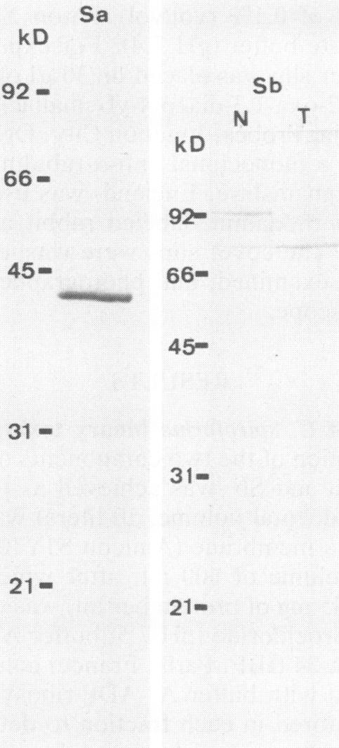
The two components Sa (Mr, 44,000) and Sb (Mr, 92,000) of Clostridium spiroforme toxin were identified and characterized. Serological data permitted the identification of two groups of actin ADP-ribosylating clostridial toxins. The first consists of only C. botulinum C2. The second group includes spiroforme toxin, iota toxin of C. perfringens E, and an enzyme called CDT found in one strain of C. difficile, antibodies against which cross-react with all of the members of both groups. C. spiroforme toxin acted on cells by disrupting microfilaments by ADP-ribosylation of G actin. Toxicity was not blocked by 10 or 20 mM ammonium chloride and was only moderately inhibited by 30 mM NH4Cl. Inhibition of coated-pit formation in HEp-2 cells by potassium depletion strongly protected against the effect of C. spiroforme toxin. Toxicity was not blocked by incubation of HEp-2 cells and spiroforme toxin at 15 degrees C. These results suggest that this new binary toxin enters cells via the coated-pit-coated-vesicle pathway and might reach the cytoplasm at the same time as or before transfer to early endosomes.
艰难梭菌毒素的两个组分Sa(分子量44,000)和Sb(分子量92,000)已被鉴定和表征。血清学数据有助于鉴定两组肌动蛋白ADP - 核糖基化梭菌毒素。第一组仅由肉毒杆菌C2型组成。第二组包括螺形菌毒素、产气荚膜梭菌E型的埃塔毒素,以及在一株艰难梭菌中发现的一种名为CDT的酶,针对该酶的抗体与两组的所有成员均发生交叉反应。螺形菌毒素通过对G肌动蛋白进行ADP - 核糖基化破坏微丝而作用于细胞。10或20 mM氯化铵不能阻断其毒性,30 mM NH4Cl仅能适度抑制其毒性。钾离子耗竭对HEp - 2细胞中被膜小窝形成的抑制作用能强烈保护细胞免受螺形菌毒素的影响。在15℃下将HEp - 2细胞与螺形菌毒素共同孵育不能阻断其毒性。这些结果表明,这种新的二元毒素通过被膜小窝 - 被膜小泡途径进入细胞,并且可能在转移至早期内体的同时或之前到达细胞质。