Bzdok Danilo, Heeger Adrian, Langner Robert, Laird Angela R, Fox Peter T, Palomero-Gallagher Nicola, Vogt Brent A, Zilles Karl, Eickhoff Simon B
Institute of Neuroscience and Medicine (INM-1), Research Center Jülich, Jülich, Germany; Institute of Clinical Neuroscience and Medical Psychology, Heinrich Heine University Düsseldorf, Düsseldorf, Germany.
Institute of Clinical Neuroscience and Medical Psychology, Heinrich Heine University Düsseldorf, Düsseldorf, Germany.
Neuroimage. 2015 Feb 1;106:55-71. doi: 10.1016/j.neuroimage.2014.11.009. Epub 2014 Nov 8.
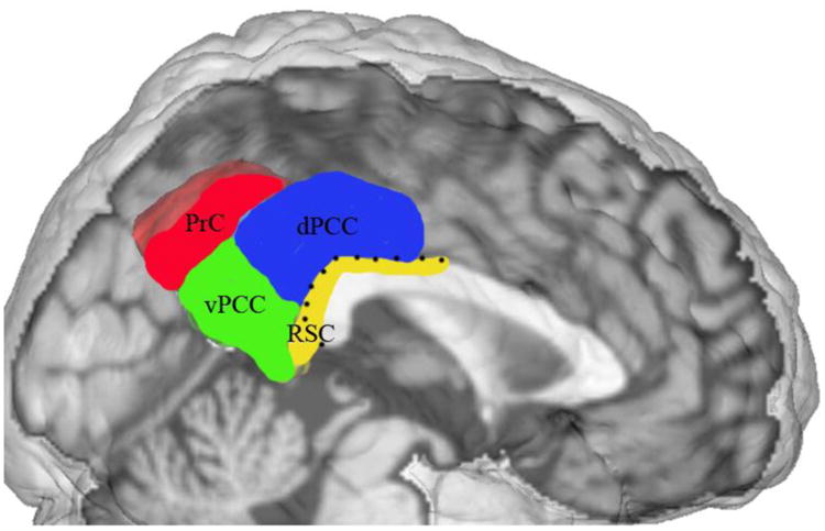
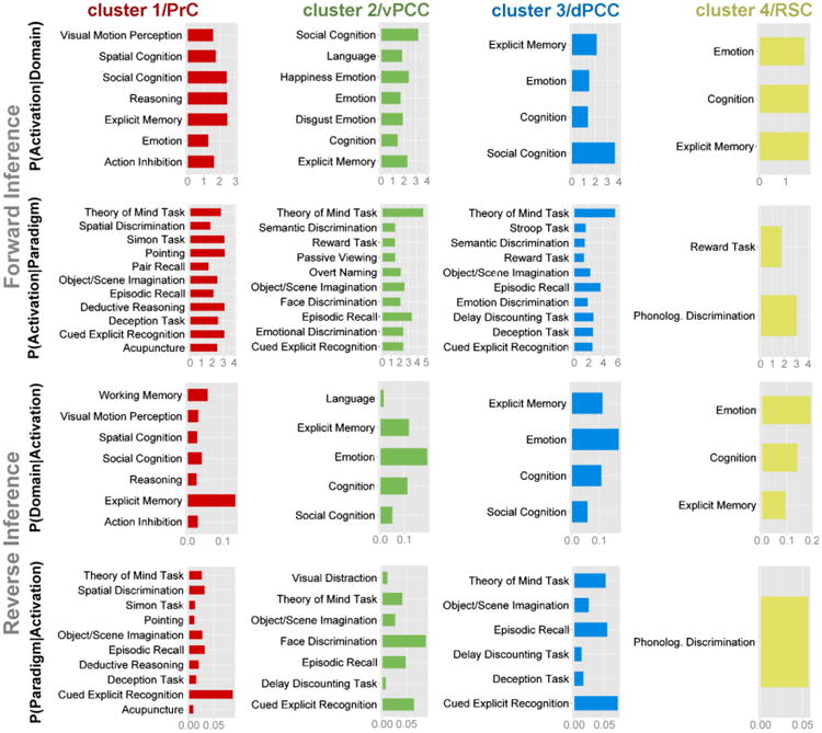
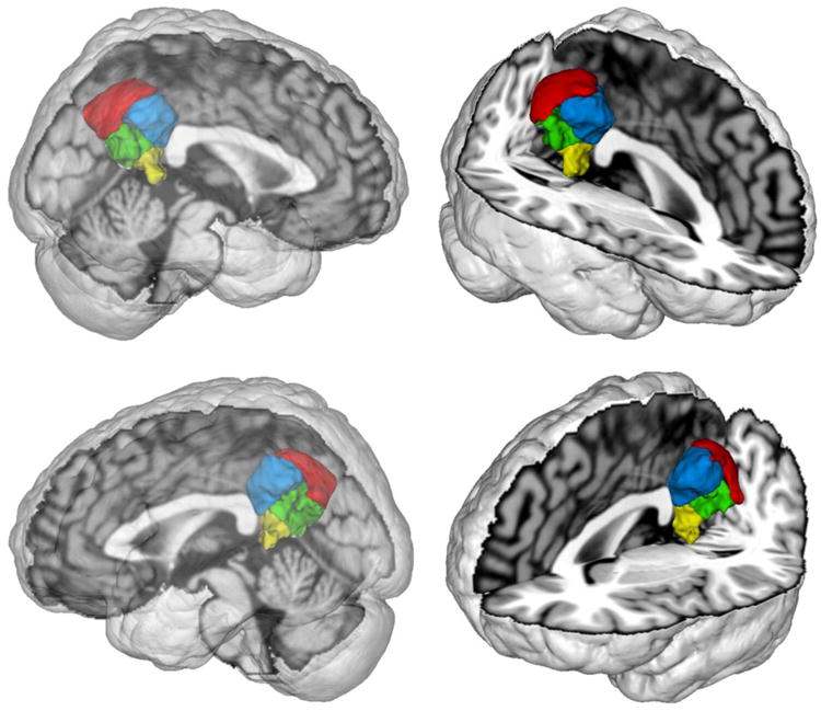
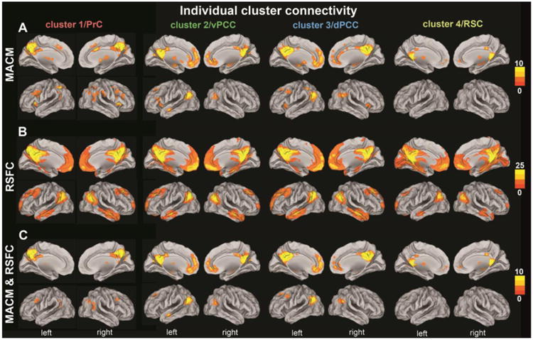
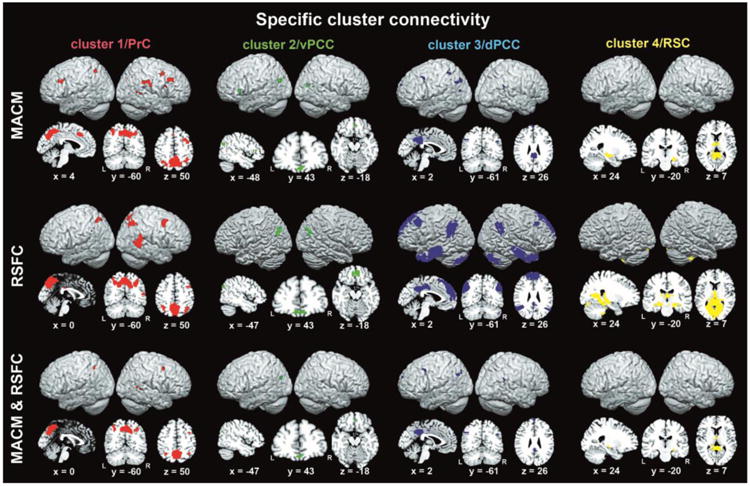
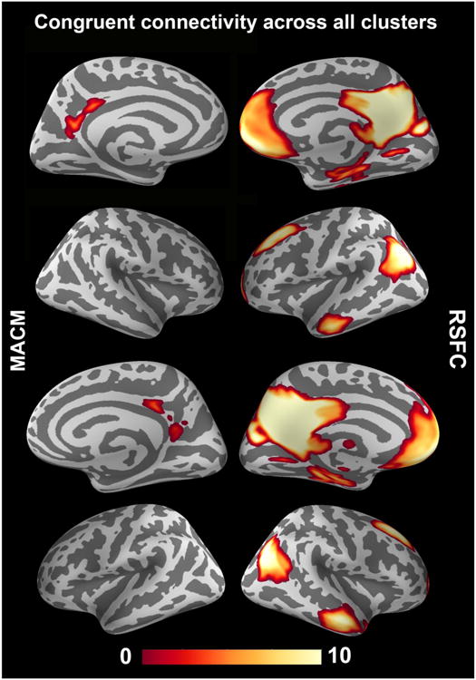
The posterior medial cortex (PMC) is particularly poorly understood. Its neural activity changes have been related to highly disparate mental processes. We therefore investigated PMC properties with a data-driven exploratory approach. First, we subdivided the PMC by whole-brain coactivation profiles. Second, functional connectivity of the ensuing PMC regions was compared by task-constrained meta-analytic coactivation mapping (MACM) and task-unconstrained resting-state correlations (RSFC). Third, PMC regions were functionally described by forward/reverse functional inference. A precuneal cluster was mostly connected to the intraparietal sulcus, frontal eye fields, and right temporo-parietal junction; associated with attention and motor tasks. A ventral posterior cingulate cortex (PCC) cluster was mostly connected to the ventromedial prefrontal cortex and middle left inferior parietal cortex (IPC); associated with facial appraisal and language tasks. A dorsal PCC cluster was mostly connected to the dorsomedial prefrontal cortex, anterior/posterior IPC, posterior midcingulate cortex, and left dorsolateral prefrontal cortex; associated with delay discounting. A cluster in the retrosplenial cortex was mostly connected to the anterior thalamus and hippocampus. Furthermore, all PMC clusters were congruently coupled with the default mode network according to task-unconstrained but not task-constrained connectivity. We thus identified distinct regions in the PMC and characterized their neural networks and functional implications.
后内侧皮质(PMC)的情况尤其鲜为人知。其神经活动变化与极为不同的心理过程有关。因此,我们采用数据驱动的探索性方法研究了PMC的特性。首先,我们通过全脑共激活图谱对PMC进行细分。其次,通过任务约束的元分析共激活映射(MACM)和任务非约束的静息态相关性(RSFC)比较了随后的PMC区域的功能连接性。第三,通过正向/反向功能推断对PMC区域进行功能描述。一个楔前叶簇主要与顶内沟、额眼区和右侧颞顶交界区相连;与注意力和运动任务相关。一个腹侧后扣带回皮质(PCC)簇主要与腹内侧前额叶皮质和左侧顶下小叶中部(IPC)相连;与面部评估和语言任务相关。一个背侧PCC簇主要与背内侧前额叶皮质、IPC的前部/后部、后扣带中部皮质和左侧背外侧前额叶皮质相连;与延迟折扣相关。一个压后皮质簇主要与丘脑前核和海马体相连。此外,根据任务非约束而非任务约束的连接性,所有PMC簇均与默认模式网络一致耦合。因此,我们在PMC中识别出了不同区域,并对其神经网络和功能意义进行了表征。