Zhuo Chuanjun, Zhu Jiajia, Qin Wen, Qu Hongru, Ma Xiaolei, Tian Hongjun, Xu Qingying, Yu Chunshui
Department of Radiology and Tianjin Key Laboratory of Functional Imaging, Tianjin Medical University General Hospital Tianjin, China ; Functional Neuroimaging Laboratory, Department of Psychiatry, Tianjin Mental Health Center, Tianjin Anding Hospital Tianjin, China ; Tianjin Anning Hospital Tianjin, China.
Department of Radiology and Tianjin Key Laboratory of Functional Imaging, Tianjin Medical University General Hospital Tianjin, China.
Front Behav Neurosci. 2014 Nov 19;8:404. doi: 10.3389/fnbeh.2014.00404. eCollection 2014.
Schizophrenia is characterized by altered resting-state functional connectivity. Most previous studies have focused on changes in connectivity strengths; however, the alterations in connectivity density in schizophrenia remain largely unknown. Here, we aimed to investigate changes in resting-state functional connectivity density (rsFCD) in schizophrenia.
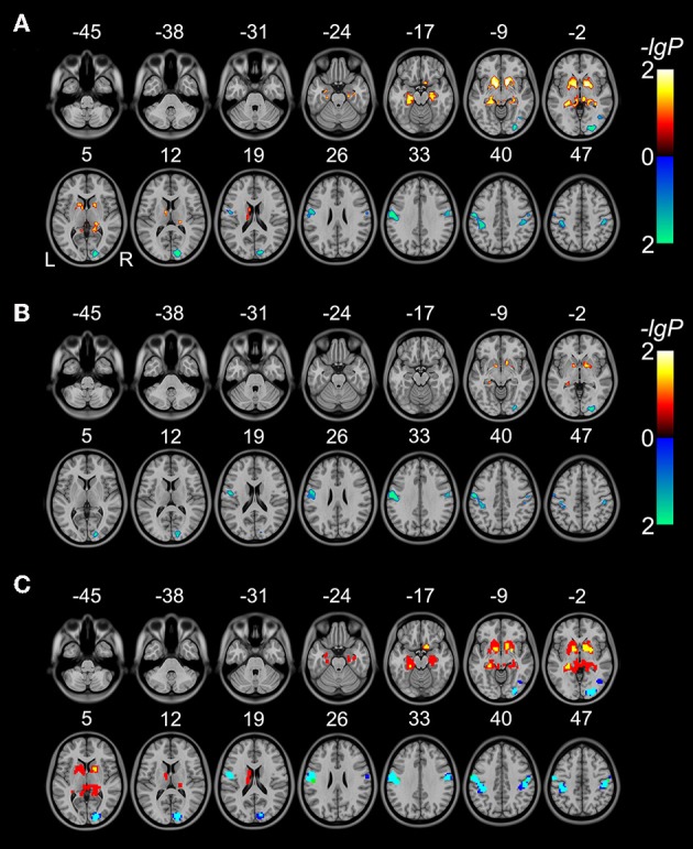
A total of 95 schizophrenia patients and 93 sex- and age-matched healthy controls (HCs) underwent resting-state functional MRI examinations. The rsFCD, which reflects the total number of functional connections between a given brain voxel and all other voxels in the entire brain, was calculated for each voxel of each subject. Voxel-based comparisons were performed to identify brain regions with significant rsFCD differences between patients and controls (P < 0.05, corrected).
Compared with HCs, patients with schizophrenia showed significantly increased rsFCD in the bilateral striatum and hippocampus and significantly decreased rsFCD in the bilateral sensorimotor cortices and right occipital cortex. However, the rsFCD values of these brain regions were not correlated with antipsychotic dosage, illness duration, or clinical symptom severity.
The striatal and hippocampal regions and parietal-occipital regions exhibited completely different changes in rsFCD in schizophrenia, which roughly correspond to dopamine activity in these regions in schizophrenia. These findings support the connectivity disorder hypothesis of schizophrenia and increase our understanding of the neural mechanisms of schizophrenia.
精神分裂症的特征是静息态功能连接改变。以往大多数研究都集中在连接强度的变化上;然而,精神分裂症中连接密度的改变在很大程度上仍不清楚。在此,我们旨在研究精神分裂症患者静息态功能连接密度(rsFCD)的变化。
共有95例精神分裂症患者和93例性别及年龄匹配的健康对照者(HCs)接受了静息态功能磁共振成像检查。计算每个受试者每个体素的rsFCD,它反映了给定脑体素与全脑所有其他体素之间功能连接的总数。进行基于体素的比较,以确定患者和对照者之间rsFCD存在显著差异的脑区(P < 0.05,校正)。
与HCs相比,精神分裂症患者双侧纹状体和海马的rsFCD显著增加,双侧感觉运动皮层和右侧枕叶皮层的rsFCD显著降低。然而,这些脑区的rsFCD值与抗精神病药物剂量、病程或临床症状严重程度无关。
精神分裂症患者纹状体和海马区域以及顶枕区域的rsFCD表现出完全不同的变化,这大致与精神分裂症中这些区域的多巴胺活性相对应。这些发现支持了精神分裂症的连接障碍假说,并增进了我们对精神分裂症神经机制的理解。