Department of Radiology and Tianjin Key Laboratory of Functional Imaging, Tianjin Medical University General Hospital, No. 154, Anshan Road, Heping District, Tianjin, 300052, China.
Department of Psychiatry, Wenzhou Seventh People's Hospital, Wenzhou, Zhejiang Province, 325000, China.
Brain Imaging Behav. 2018 Apr;12(2):383-389. doi: 10.1007/s11682-017-9704-0.
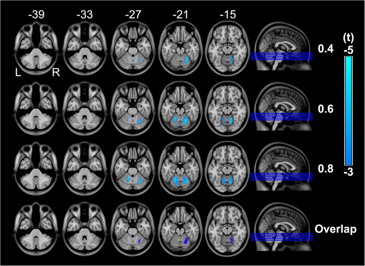
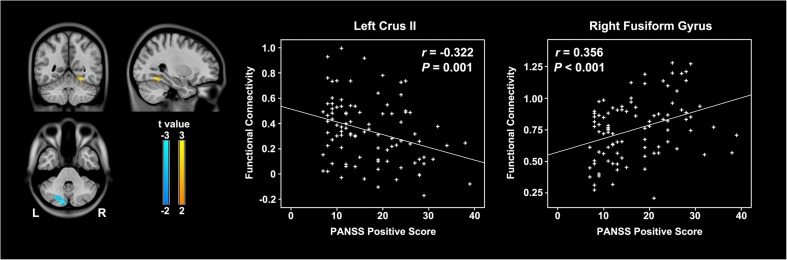
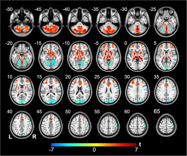
Structural and functional abnormalities of the cerebellum in schizophrenia have been reported. Most previous studies investigating resting-state functional connectivity (rsFC) have relied on a priori restrictions on seed regions or specific networks, which may bias observations. In this study, we aimed to elicit the connectivity alterations of the cerebellum in schizophrenia in a hypothesis-free approach. Ninety-five schizophrenia patients and 93 sex- and age-matched healthy controls underwent resting-state functional magnetic resonance imaging (fMRI). A voxel-wise data-driven method, resting-state functional connectivity density (rsFCD), was used to investigate cerebellar connectivity changes in schizophrenia patients. Regions with altered rsFCD were chosen as seeds to perform seed-based resting-state functional connectivity (rsFC) analyses. We found that schizophrenia patients exhibited decreased rsFCD in the right hemispheric VI; moreover, this cerebellar region showed increased rsFC with the prefrontal cortex and subcortical nuclei and decreased rsFC with the visual cortex and sensorimotor cortex. In addition, some rsFC changes were associated with positive symptoms. These findings suggest that abnormalities of the cerebellar hub and cerebellar-subcortical-cortical loop may be the underlying mechanisms of schizophrenia.
已有研究报道精神分裂症患者小脑结构和功能异常。既往大多数研究采用基于先验假设的种子区域或特定网络来探究静息态功能连接(rsFC),这可能会导致观察结果存在偏倚。本研究旨在采用无假设的方法来探究精神分裂症患者小脑的连接变化。95 例精神分裂症患者和 93 名性别和年龄匹配的健康对照者接受了静息态功能磁共振成像(fMRI)检查。采用基于体素的静息态功能连接密度(rsFCD)数据驱动方法,来研究精神分裂症患者小脑的连接变化。选择具有改变的 rsFCD 的区域作为种子点进行基于种子的静息态功能连接(rsFC)分析。我们发现,精神分裂症患者右侧小脑 VI 区域的 rsFCD 降低;此外,该小脑区域与前额叶皮层和皮质下核团的 rsFC 增加,与视觉皮层和感觉运动皮层的 rsFC 降低。此外,一些 rsFC 的变化与阳性症状有关。这些发现表明,小脑中枢和小脑-皮质下-皮层环路的异常可能是精神分裂症的潜在机制。