Chen Jianying, Liu Zhenjun, Hong Mian Ming, Zhang Hongzhe, Chen Can, Xiao Mengyuan, Wang Junxian, Yao Feng, Ba Mingchuan, Liu Jinghu, Guo Zi-Kuan, Zhong Jixin
Division of Cardiovascular Disease, Department of Internal Medicine, Affiliated Hospital of Guangdong Medical College, Zhanjiang, Guangdong, China.
Department of Experimental Hematology, Beijing Institute of Radiation Medicine, Beijing, China.
PLoS One. 2014 Dec 16;9(12):e115316. doi: 10.1371/journal.pone.0115316. eCollection 2014.
INTRODUCTION & OBJECTIVE: Microvesicles (MVs) derived from mesenchymal stem cells (MSCs) have been shown to promote angiogenesis. This study was aimed to shed a light on the mechanisms by analyzing the angiogenesis-promoting compositions of MSC-MVs. Also we try to figure out the impact of hypoxia on angiogenesis.
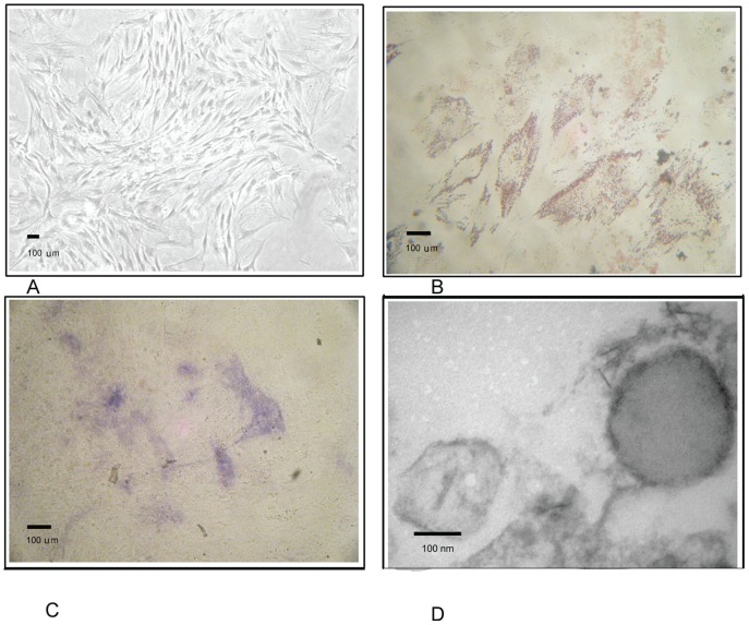
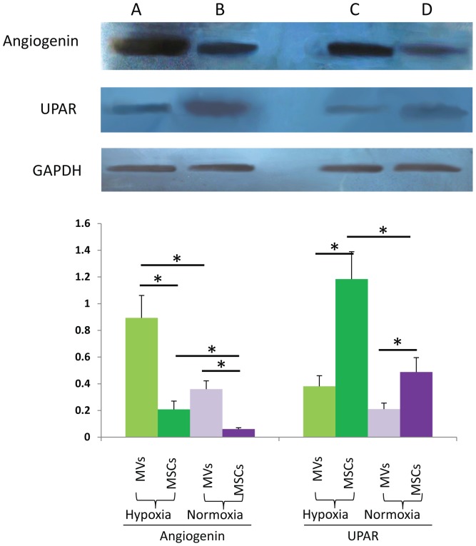
MVs were isolated from the culture supernatants of MSCs under hypoxia/normoxia and serum-deprivation condition. The morphological features of MVs were revealed by an electron microscope and the origin of the MVs was identified by a bead-bound assay. An antibody array was used to analyze the expression of angiogenic cytokines from MVs and the parent MSCs as well. The major candidate factors were screened and the results were validated by immune blotting.
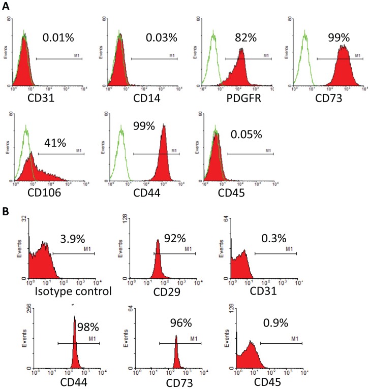
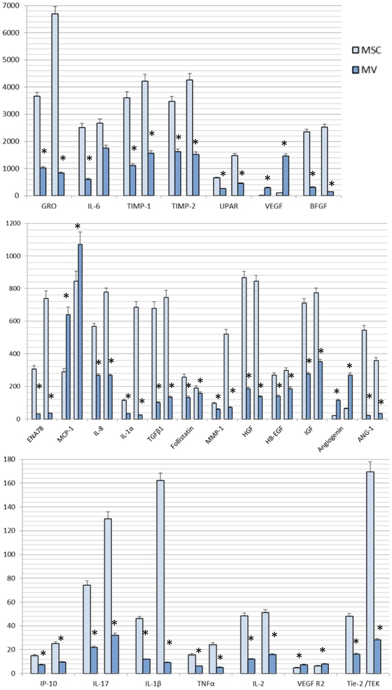
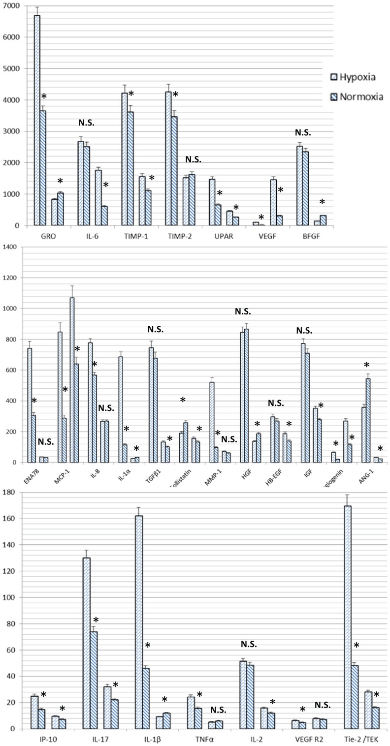
MSC-MVs were around 80 nm in diameter. They expressed CD29, CD44, and CD73, but not CD31 and CD45. Antibody array showed that both MSCs and MVs expressed many angiogenesis-promoting biomolecules, including interleukin-6 (IL-6), basic fibroblast growth factors (bFGF), and recptor of urokinase-type plasminogen activator (UPAR). MSC-MVs contained angiogenin, vascular endothelial growth factor (VEGF), monocyte chemotactic protein-1 (MCP-1) and the receptor-2 for vascular endothelial growth factor at higher levels than the parent MSCs. Under hypoxic condition most cytokines were expressed in greater quantity than normoxic in MSCs while in MVs there was no significant difference between hypoxic and normoxic conditions except UPAR, Angiogenin, VEGF, IGF, Tie-2/TEK, and IL-6 which were higher in MVs under hypoxic conditions than those in normoxic condition.
Upon serum-deprivation condition, MSCs could secrete MVs that contain a variety of factors contributing to their angiogenesis-promoting function. And among them, Angiogenin, VEGF, MCP-1, VEGF R2 might be of greater importance than the other cytokines. Also UPAR, Angiogenin, VEGF, IGF, Tie-2/TEK, IL-6 might be responsible for hypoxia-augmented proangiogenic effects of MVs.
间充质干细胞(MSC)来源的微泡(MV)已被证明可促进血管生成。本研究旨在通过分析MSC-MV的促血管生成成分来阐明其机制。此外,我们试图弄清楚缺氧对血管生成的影响。
在缺氧/常氧和血清饥饿条件下,从MSC的培养上清液中分离MV。通过电子显微镜揭示MV的形态特征,并通过磁珠结合试验鉴定MV的来源。使用抗体芯片分析MV和原始MSC中血管生成细胞因子的表达。筛选主要候选因子,并通过免疫印迹验证结果。
MSC-MV直径约为80nm。它们表达CD29、CD44和CD73,但不表达CD31和CD45。抗体芯片显示,MSC和MV均表达许多促血管生成生物分子,包括白细胞介素-6(IL-6)、碱性成纤维细胞生长因子(bFGF)和尿激酶型纤溶酶原激活剂受体(UPAR)。MSC-MV中血管生成素、血管内皮生长因子(VEGF)、单核细胞趋化蛋白-1(MCP-1)和血管内皮生长因子受体-2的含量高于原始MSC。在缺氧条件下,MSC中大多数细胞因子的表达量比常氧时更高,而在MV中,除了UPAR、血管生成素、VEGF、胰岛素样生长因子(IGF)、酪氨酸激酶受体2/TEK(Tie-2/TEK)和IL-6在缺氧条件下的表达量高于常氧条件外,缺氧和常氧条件下没有显著差异。
在血清饥饿条件下,MSC可分泌含有多种促进血管生成功能因子的MV。其中,血管生成素、VEGF、MCP-1、VEGF R2可能比其他细胞因子更重要。此外,UPAR、血管生成素、VEGF、IGF、Tie-2/TEK、IL-6可能是MV缺氧增强促血管生成作用的原因。