Nielsen Flemming K, Egund Niels, Peters David, Jurik Anne Grethe
Department of Radiology, Aarhus University Hospital, Noerrebrogade 44, 8000 Aarhus, Denmark.
BMC Musculoskelet Disord. 2014 Dec 20;15:447. doi: 10.1186/1471-2474-15-447.
Longitudinal assessment of bone marrow lesions (BMLs) in knee osteoarthritis (KOA) by MRI is usually performed using semi-quantitative grading methods. Quantitative segmentation methods may be more sensitive to detect change over time. The purpose of this study was to evaluate and compare the validity and sensitivity to detect changes of two quantitative MR segmentation methods for measuring BMLs in KOA, one computer assisted (CAS) and one manual (MS) method.
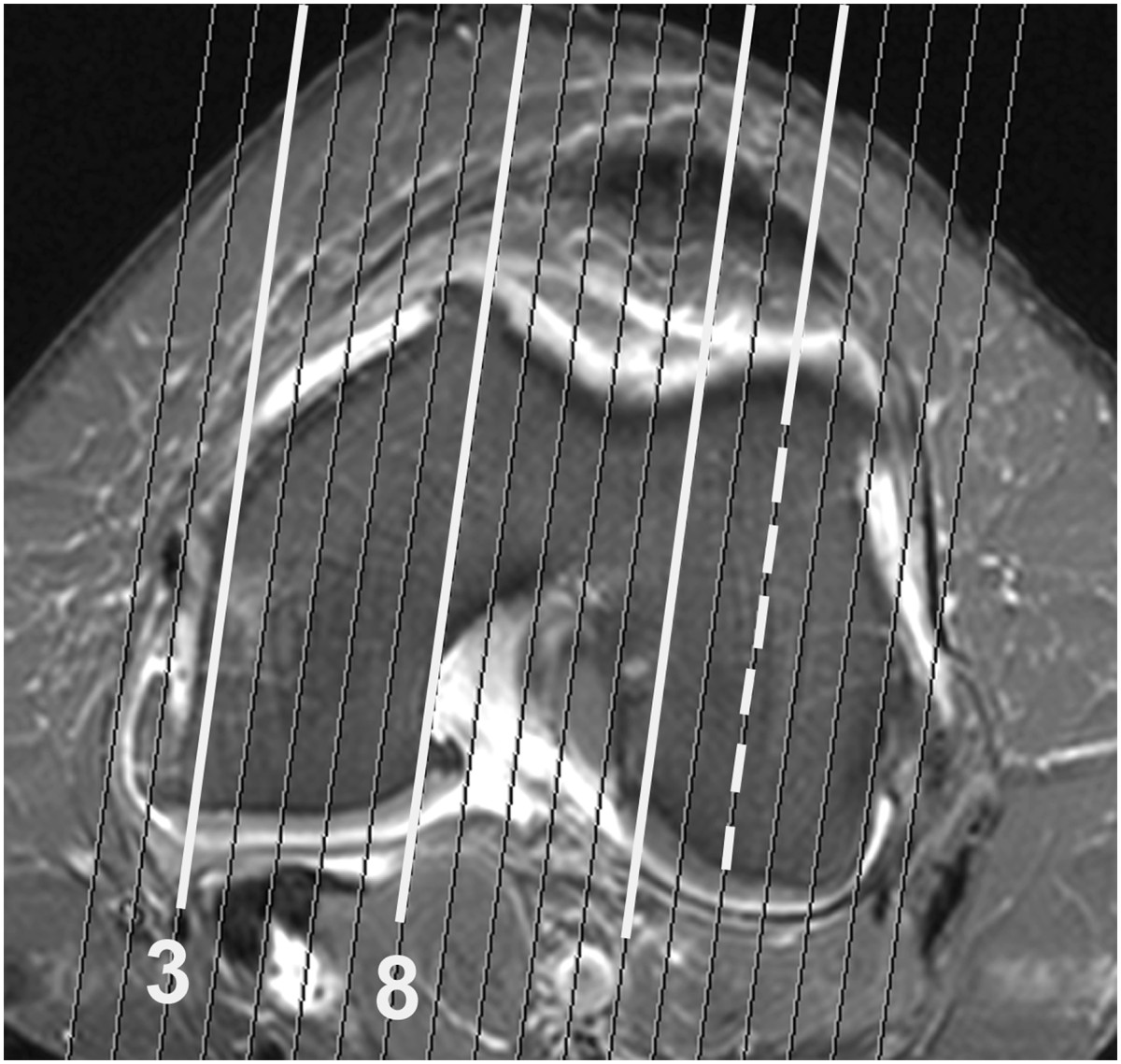
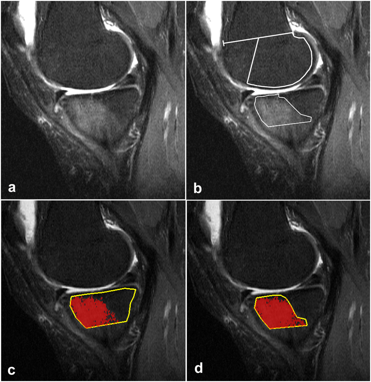
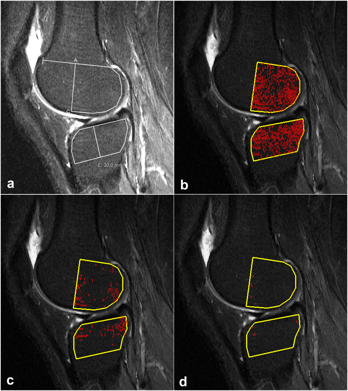
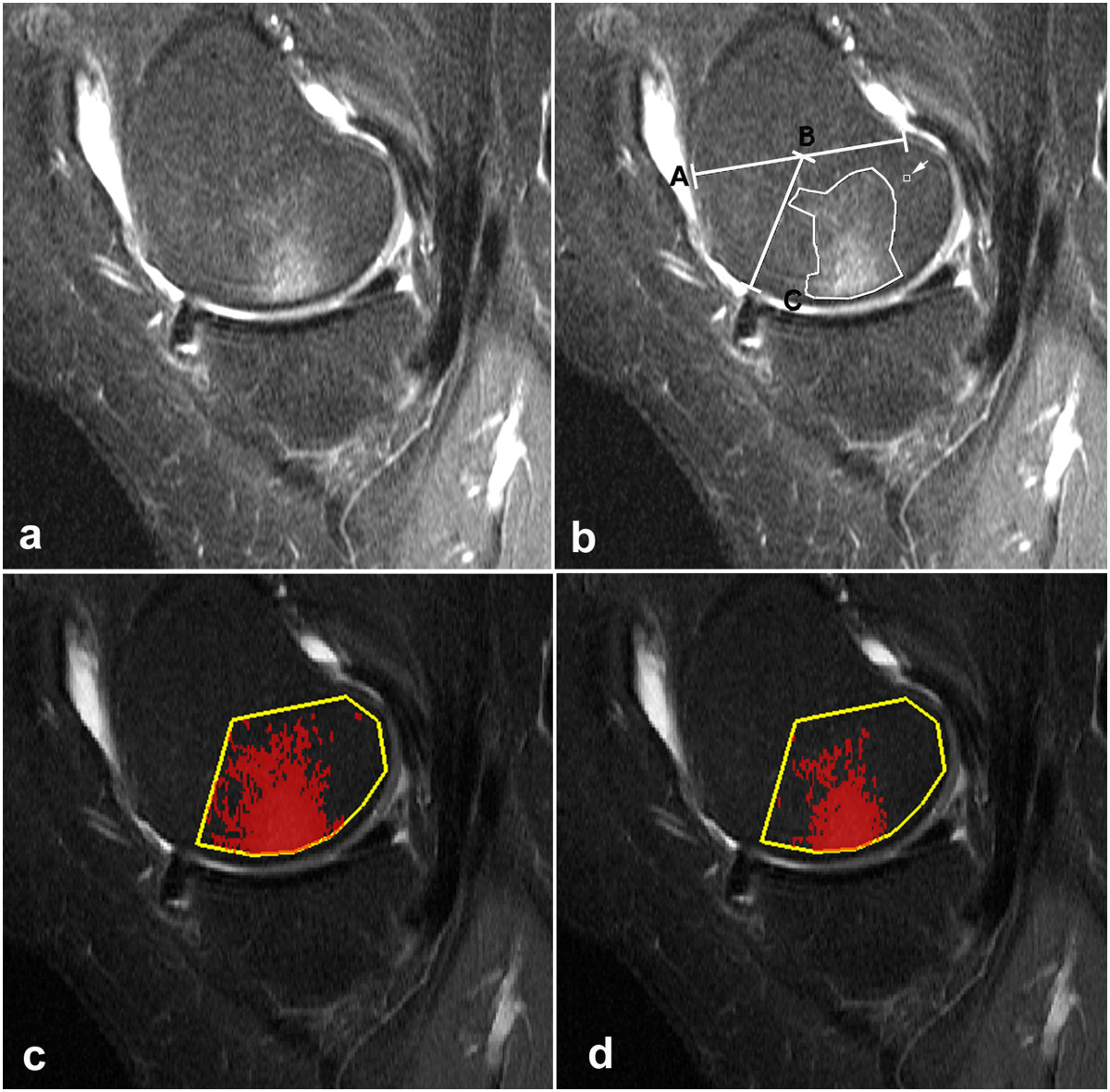
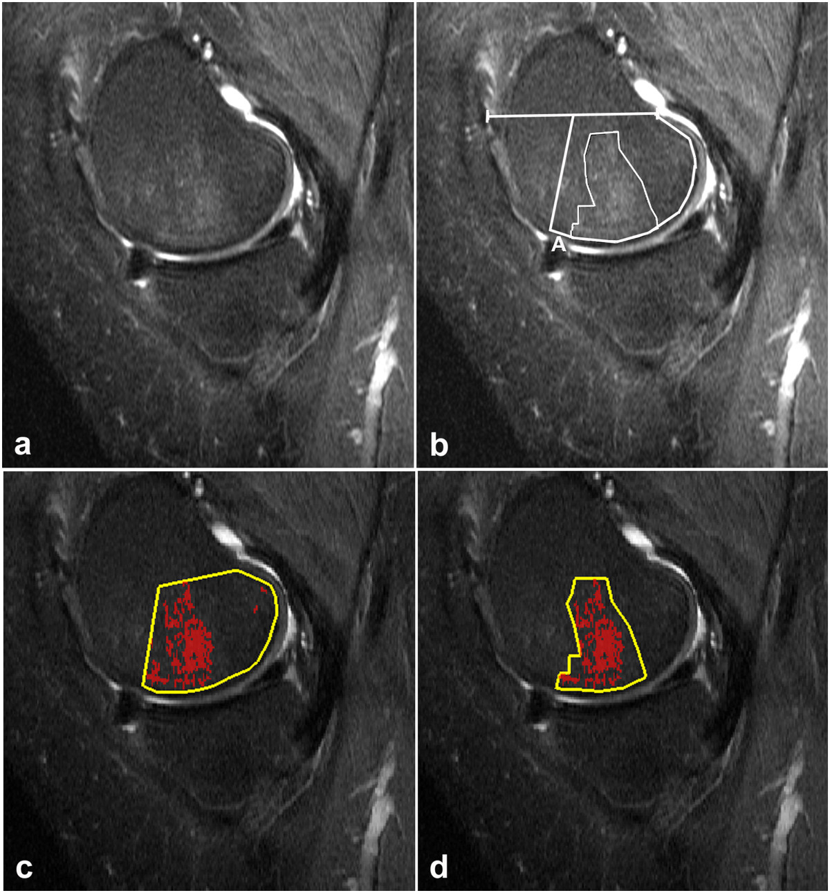
Twenty-two patients with KOA confined to the medial femoro-tibial compartment obtained MRI at baseline and follow-up (median 334 days in between). STIR, T1 and fat saturated T1 post-contrast sequences were obtained using a 1.5 T system. The 44 sagittal STIR sequences were assessed independently by two readers for quantification of BML. The signal intensities (SIs) of the normal bone marrow in the lateral femoral condyles and tibial plateaus were used as threshold values. The volume of bone marrow with SIs exceeding the threshold values (BML) was measured in the medial femoral condyle and tibial plateau and related to the total volume of the condyles/plateaus.The 95% limits of agreement at baseline were used to determine the sensitivity to change.
The mean threshold values of CAS and MS were almost identical but the absolute and relative BML volumes differed being 1319 mm3/10% and 1828 mm3/15% in the femur and 941 mm3/7% and 2097 mm3/18% in the tibia using CAS and MS, respectively. The BML volumes obtained by CAS and MS were significantly correlated but the tissue changes measured were different. The volume of voxels exceeding the threshold values was measured by CAS whereas MS included intervening voxels with normal SI.The 95% limits of agreement were narrower by CAS than by MS; a significant change of relative BML by CAS was outside the limits of -2.0%-4.7% whereas the limits by MS were -6.9%-8.2%. The BML changed significantly in 13 knees using CAS and in 10 knees by MS.
CAS was a reliable method for measuring BML and more sensitive to detect changes over time than MS. The BML volumes measured by the two methods differed but were significantly correlated.
膝关节骨关节炎(KOA)中骨髓损伤(BMLs)的纵向MRI评估通常采用半定量分级方法。定量分割方法在检测随时间的变化方面可能更敏感。本研究的目的是评估和比较两种用于测量KOA中BMLs的定量MR分割方法(一种计算机辅助(CAS)方法和一种手动(MS)方法)检测变化的有效性和敏感性。
22例局限于股骨内侧髁间室的KOA患者在基线和随访时(间隔中位数为334天)接受了MRI检查。使用1.5T系统获取STIR、T1和脂肪饱和T1增强后序列。44个矢状面STIR序列由两名阅片者独立评估以量化BML。以外侧股骨髁和胫骨平台正常骨髓的信号强度(SIs)作为阈值。测量股骨内侧髁和胫骨平台中SI超过阈值的骨髓体积(BML),并与髁/平台的总体积相关。基线时的95%一致性界限用于确定对变化的敏感性。
CAS和MS的平均阈值几乎相同,但绝对和相对BML体积不同,使用CAS和MS时,股骨中的分别为1319mm³/10%和1828mm³/15%,胫骨中的分别为941mm³/7%和2097mm³/18%。CAS和MS获得的BML体积显著相关,但测量的组织变化不同。CAS测量的是超过阈值的体素体积,而MS包括具有正常SI的中间体素。CAS的95%一致性界限比MS更窄;CAS测量的相对BML的显著变化超出了-2.0%-4.7%的界限,而MS的界限为-6.9%-8.2%。使用CAS时13个膝关节的BML有显著变化,使用MS时10个膝关节的BML有显著变化。
CAS是一种可靠的测量BML的方法,并且比MS更敏感地检测随时间的变化。两种方法测量的BML体积不同,但显著相关。