Ocak Meltem, Gillman Andrea G, Bresee Jamee, Zhang Lixin, Vlad Anda M, Müller Cristina, Schibli Roger, Edwards W Barry, Anderson Carolyn J, Gach H Michael
Department of Radiology, ‡Cancer Institute, §Department of Obstetrics, Gynecology & Reproductive Sciences, ∥Magee Womens Research Institute, ⊥Department of Pharmacology & Chemical Biology, #Department of Bioengineering, University of Pittsburgh , Pittsburgh, Pennsylvania 15213, United States.
Mol Pharm. 2015 Feb 2;12(2):542-53. doi: 10.1021/mp500628g. Epub 2015 Jan 14.
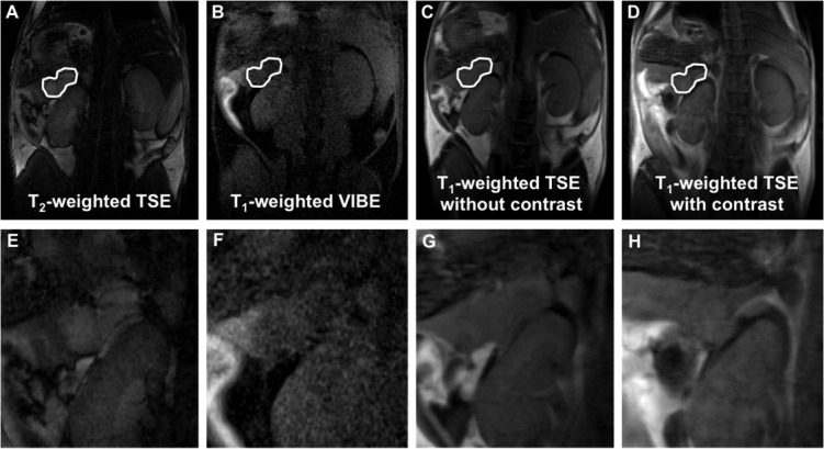
A new transplantable ovarian tumor model is presented using a novel folate receptor (FR) positive, murine ovarian cancer cell line that emulates the human disease and induces widespread intraperitoneal (i.p.) tumors in immunocompetent mice within 4-8 weeks of implantation. Tumor development was monitored using a new positron emission tomography (PET) FR-targeting reporter with PET/computerized tomography (PET/CT) and fluorescence molecular tomography (FMT) using a commercial FR-targeting reporter. Conventional structural magnetic resonance imaging (MRI) was also performed. Adult female C57BL/6 mice were injected i.p. with 6 × 10(6) MKP-L FR+ cells. Imaging was performed weekly beginning 2 weeks after tumor induction. The albumin-binding, FR-targeting ligand cm09 was radiolabeled with the positron emitter (68)Ga and used to image the tumors with a small animal PET/CT. The FR-reporter FolateRSense 680 (PerkinElmer) was used for FMT and flow cytometry. Preclinical MRI (7 T) without FR-targeting was compared with the PET and FMT molecular imaging. Tumors were visible by all three imaging modalities. PET/CT had the highest imaging sensitivity at 3-3.5 h postadministration (mean %IA/g mean > 6) and visualized tumors earlier than the other two modalities with lower kidney uptake (mean %IA/g mean < 17) than previously reported FR-targeting agents in late stage disease. FMT showed relatively low FR-targeted agent in the bladder and kidneys, but yielded the lowest anatomical image resolution. MRI produced the highest resolution images, but it was difficult to distinguish tumors from abdominal organs during early progression since a FR-targeting MRI reporter was not used. Nevertheless, there was good correlation of imaging biomarkers between the three modalities. Tumors in the mouse ovarian cancer model could be detected using FR-targeted imaging as early as 2 weeks post i.p. injection of tumor cells. An imaging protocol should combine one or more of the modalities, e.g., PET/CT or PET/MRI for optimal tumor detection and delineation from surrounding tissues.
本文介绍了一种新的可移植性卵巢肿瘤模型,该模型使用一种新型的叶酸受体(FR)阳性小鼠卵巢癌细胞系,其可模拟人类疾病,并在免疫活性小鼠植入后4 - 8周内诱发广泛的腹腔内(i.p.)肿瘤。使用一种新的正电子发射断层扫描(PET)FR靶向报告分子,结合PET/计算机断层扫描(PET/CT)和使用商业FR靶向报告分子的荧光分子断层扫描(FMT)来监测肿瘤的发展。还进行了传统的结构磁共振成像(MRI)。成年雌性C57BL/6小鼠腹腔注射6×10(6) MKP-L FR+细胞。从肿瘤诱导后2周开始每周进行成像。白蛋白结合的FR靶向配体cm09用正电子发射体(68)Ga进行放射性标记,并用于小动物PET/CT对肿瘤进行成像。FR报告分子FolateRSense 680(PerkinElmer)用于FMT和流式细胞术。将无FR靶向的临床前MRI(7 T)与PET和FMT分子成像进行比较。所有三种成像方式均能检测到肿瘤。PET/CT在给药后3 - 3.5小时具有最高的成像灵敏度(平均%IA/g平均值> 6),并且比其他两种方式更早地可视化肿瘤,且肾脏摄取较低(平均%IA/g平均值< 17),低于先前报道的晚期疾病中的FR靶向剂。FMT显示膀胱和肾脏中的FR靶向剂相对较少,但产生的解剖图像分辨率最低。MRI产生的图像分辨率最高,但由于未使用FR靶向MRI报告分子,在早期进展过程中难以将肿瘤与腹部器官区分开来。尽管如此,三种方式之间的成像生物标志物具有良好的相关性。在小鼠卵巢癌模型中,早在腹腔注射肿瘤细胞后2周就可以使用FR靶向成像检测到肿瘤。成像方案应结合一种或多种方式,例如PET/CT或PET/MRI,以实现最佳的肿瘤检测以及与周围组织的区分。