Możdżeń Edyta, Kajta Małgorzata, Wąsik Agnieszka, Lenda Tomasz, Antkiewicz-Michaluk Lucyna
Department of Neurochemistry, Institute of Pharmacology Polish Academy of Sciences, 12 Smętna Street, 31-343, Kraków, Poland,
Neurotox Res. 2015 Apr;27(3):300-13. doi: 10.1007/s12640-014-9511-y. Epub 2014 Dec 24.
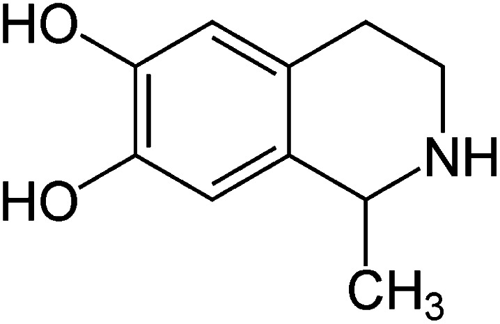
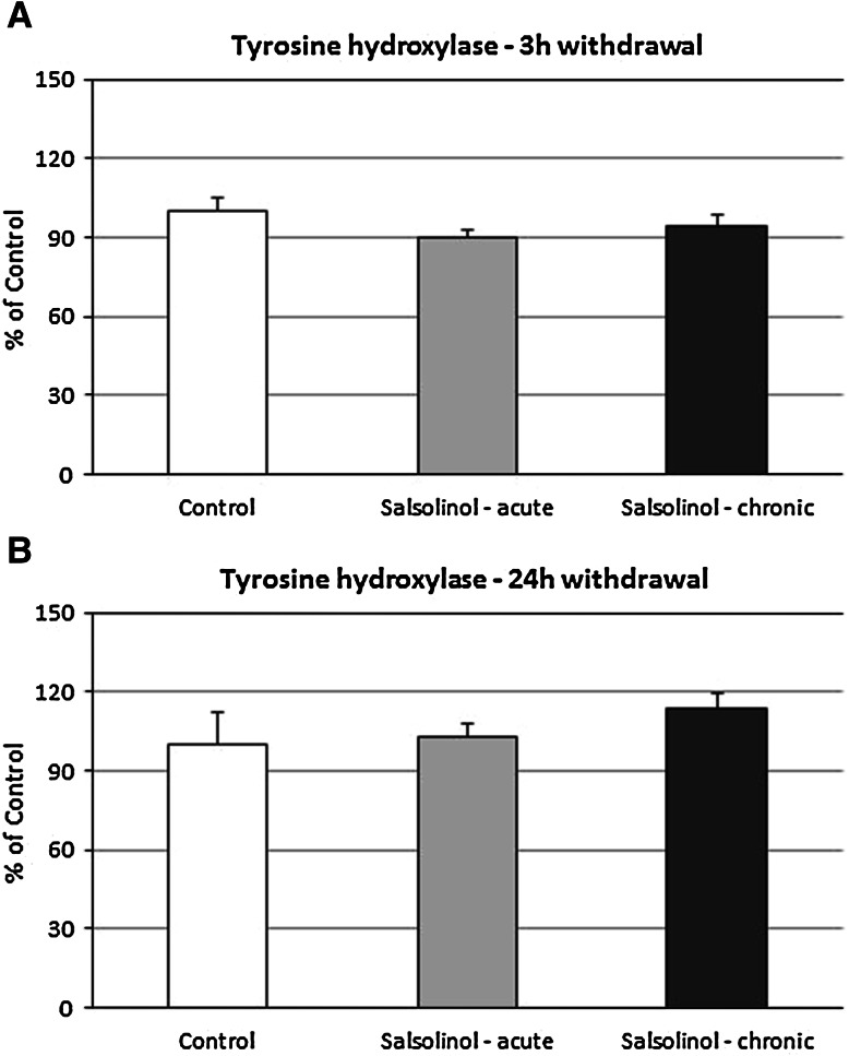
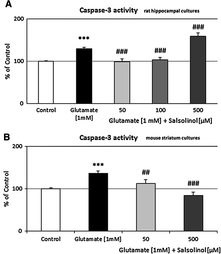
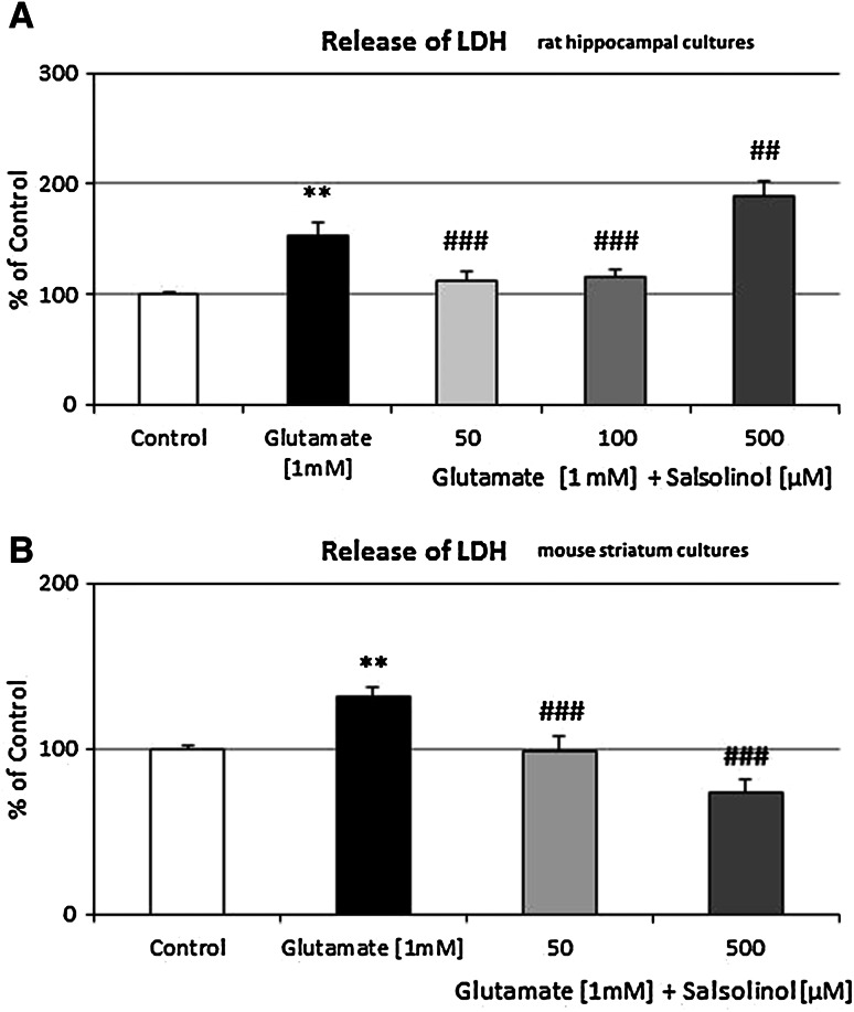
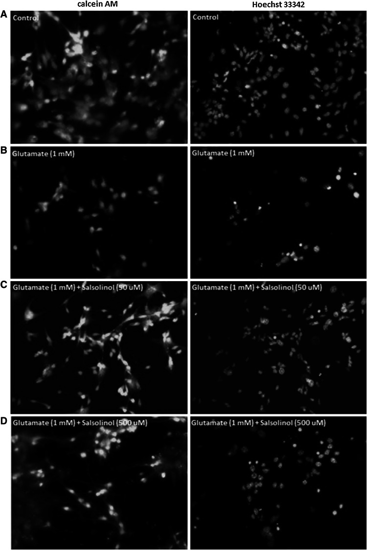
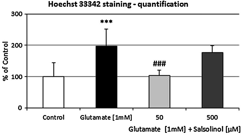
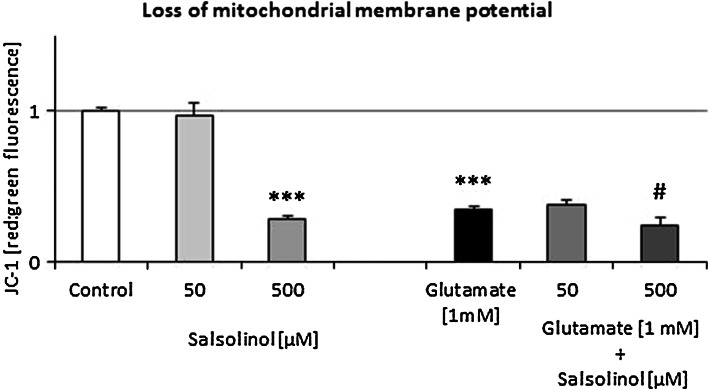
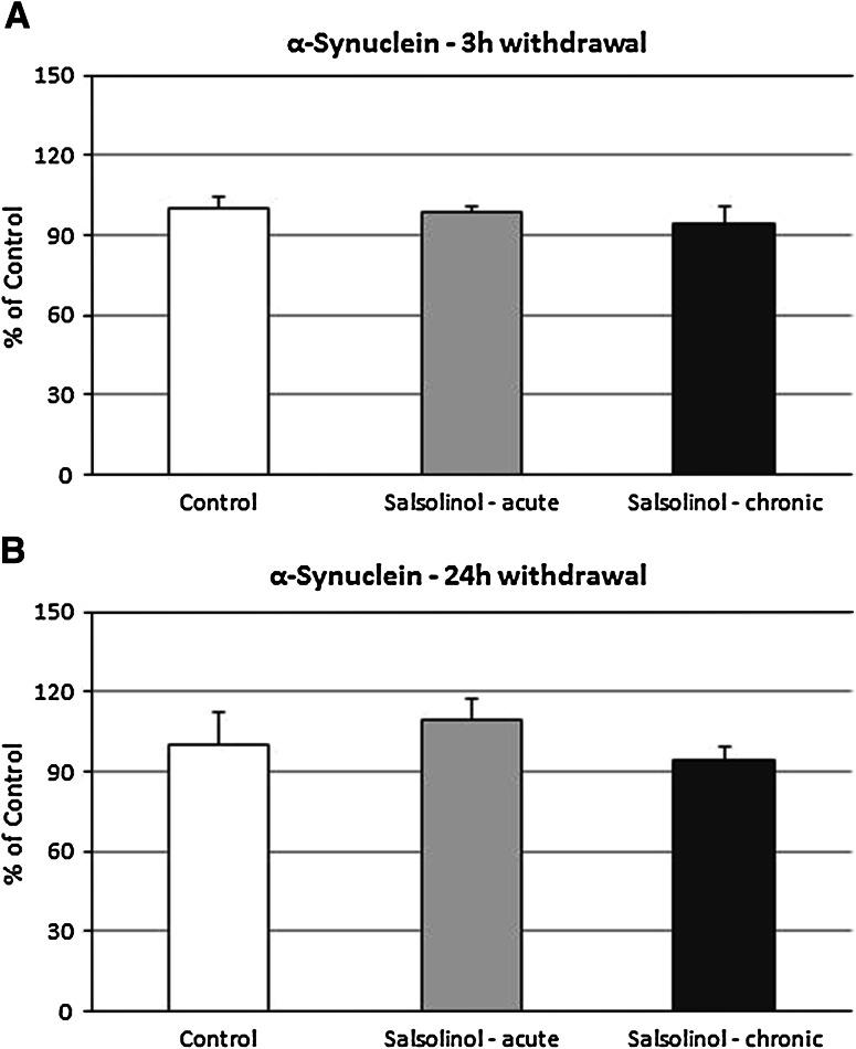
Salsolinol (1-methyl-6,7-dihydroxy-1,2,3,4-tetrahydroisoquinoline), an endogenous compound present in the brain, was suspected of participation in the etiopathogenesis of Parkinson's disease, the most common serious movement disorder worldwide. In this study, we evaluated the effect of different (50, 100, and 500 µM) concentrations of salsolinol on markers of glutamate-induced apoptotic and neurotoxic cell damage, such as caspase-3 activity, lactate dehydrogenase (LDH) release, and the loss of mitochondrial membrane potential. Biochemical data were complemented with the cellular analysis, including Hoechst 33342 and calcein AM staining, to visualize apoptotic DNA-fragmentation and to assess cell survival, respectively. The assessment of all investigated parameters was performed in primary cultures of rat or mouse hippocampal and striatum cells. Our study showed that salsolinol had biphasic effects, namely, at lower concentrations (50 and 100 µM), it demonstrated a distinct neuroprotective activity, whereas in the highest one (500 µM) caused neurotoxic effect. Salsolinol in concentrations of 50 and 100 µM significantly antagonized the pro-apoptotic and neurotoxic effects caused by 1 mM glutamate. Salsolinol diminished the number of bright fragmented nuclei with condensed chromatin and increased cell survival in Hoechst 33342 and calcein AM staining in hippocampal cultures. Additionally, in the low 50 µM concentration, it produced a significant inhibition of glutamate-induced loss of membrane mitochondrial potential. Only the highest concentration of salsolinol (500 µM) enhanced the glutamate excitotoxicity. Ex vivo studies indicated that both acute and chronic administration of salsolinol did not affect the dopamine metabolism, its striatal concentration or α-synuclein and tyrosine hydroxylase protein level in the rat substantia nigra and striatum. Summarizing, the present studies exclude possibility that salsolinol under physiological conditions could be an endogenous factor involved in the neurogenerative processes; conversely, it can exert a protective action on nerve cells in the brain. These findings may have important implications for the development of the new strategies to treat or prevent neural degeneration.
盐索诺醇(1-甲基-6,7-二羟基-1,2,3,4-四氢异喹啉)是一种存在于大脑中的内源性化合物,被怀疑参与了帕金森病的发病机制,帕金森病是全球最常见的严重运动障碍。在本研究中,我们评估了不同浓度(50、100和500μM)的盐索诺醇对谷氨酸诱导的凋亡和神经毒性细胞损伤标志物的影响,如半胱天冬酶-3活性、乳酸脱氢酶(LDH)释放以及线粒体膜电位的丧失。生化数据通过细胞分析得到补充,包括Hoechst 33342和钙黄绿素AM染色,分别用于观察凋亡DNA片段化和评估细胞存活情况。所有研究参数的评估均在大鼠或小鼠海马和纹状体细胞的原代培养物中进行。我们的研究表明,盐索诺醇具有双相作用,即在较低浓度(50和100μM)时,它表现出明显的神经保护活性,而在最高浓度(500μM)时则产生神经毒性作用。50和100μM浓度的盐索诺醇显著拮抗了1 mM谷氨酸引起的促凋亡和神经毒性作用。盐索诺醇减少了海马培养物中染色质浓缩的明亮碎片化细胞核数量,并增加了Hoechst 33342和钙黄绿素AM染色中的细胞存活。此外,在50μM的低浓度下,它对谷氨酸诱导的线粒体膜电位丧失产生了显著抑制作用。只有最高浓度的盐索诺醇(500μM)增强了谷氨酸兴奋性毒性。体内研究表明,急性和慢性给予盐索诺醇均不影响大鼠黑质和纹状体中的多巴胺代谢、其纹状体浓度或α-突触核蛋白和酪氨酸羟化酶蛋白水平。总之,本研究排除了盐索诺醇在生理条件下可能是参与神经退行性过程的内源性因素这一可能性;相反,它可以对大脑中的神经细胞发挥保护作用。这些发现可能对开发治疗或预防神经退行性变的新策略具有重要意义。