Andalusian Center for Molecular Biology and Regenerative Medicine (CABIMER), University Pablo de Olavide, Biomedical Research Network (CIBER) of Diabetes and Related Metabolic Diseases, Red-Tercel, Avenida Américo Vespucio S/N, 41092 Seville, Spain ; Sanford Consortium for Regenerative Medicine, University of California San Diego, 2880 Torrey Pines Scenic Drive, La Jolla, CA 92037, USA.
Andalusian Center for Molecular Biology and Regenerative Medicine (CABIMER), University Pablo de Olavide, Biomedical Research Network (CIBER) of Diabetes and Related Metabolic Diseases, Red-Tercel, Avenida Américo Vespucio S/N, 41092 Seville, Spain.
Stem Cells Int. 2014;2014:379678. doi: 10.1155/2014/379678. Epub 2014 Dec 3.
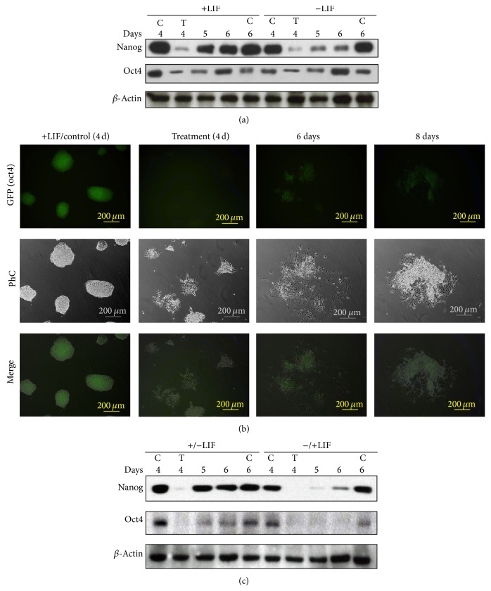
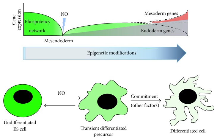
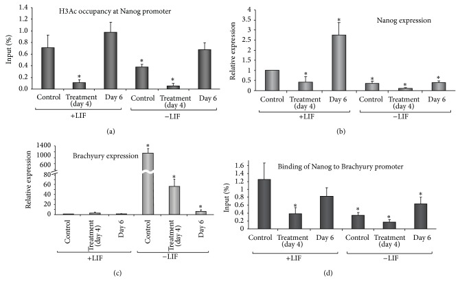
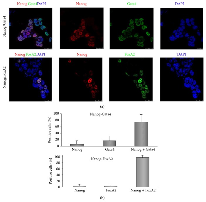
The function of pluripotency genes in differentiation is a matter of investigation. We report here that Nanog and Oct4 are reexpressed in two mouse embryonic stem cell (mESC) lines following exposure to the differentiating agent DETA/NO. Both cell lines express a battery of both endoderm and mesoderm markers following induction of differentiation with DETA/NO-based protocols. Confocal analysis of cells undergoing directed differentiation shows that the majority of cells expressing Nanog express also endoderm genes such as Gata4 and FoxA2 (75.4% and 96.2%, resp.). Simultaneously, mRNA of mesodermal markers Flk1 and Mef2c are also regulated by the treatment. Acetylated histone H3 occupancy at the promoter of Nanog is involved in the process of reexpression. Furthermore, Nanog binding to the promoter of Brachyury leads to repression of this gene, thus disrupting mesendoderm transition.
多能性基因在分化中的功能是一个研究的问题。我们在这里报告,在暴露于分化剂 DETA/NO 后,两种小鼠胚胎干细胞(mESC)系中重新表达了 Nanog 和 Oct4。在使用基于 DETA/NO 的方案诱导分化后,这两个细胞系都表达了一系列内胚层和中胚层标记物。对定向分化细胞进行共聚焦分析表明,大多数表达 Nanog 的细胞也表达内胚层基因,如 Gata4 和 FoxA2(分别为 75.4%和 96.2%)。同时,中胚层标记物 Flk1 和 Mef2c 的 mRNA 也受到该处理的调节。组蛋白 H3 在 Nanog 启动子上的乙酰化占据参与了重新表达的过程。此外,Nanog 与 Brachyury 启动子结合导致该基因的抑制,从而破坏中胚层-内胚层过渡。