1. College of Oriental Medicine, Kyung Hee University, Seoul 130-701, South Korea.
2. Graduate School of East-West Medical Science, Kyung Hee University, Yongin 449-701, Republic of Korea.
J Cancer. 2015 Jan 1;6(1):19-28. doi: 10.7150/jca.9591. eCollection 2015.
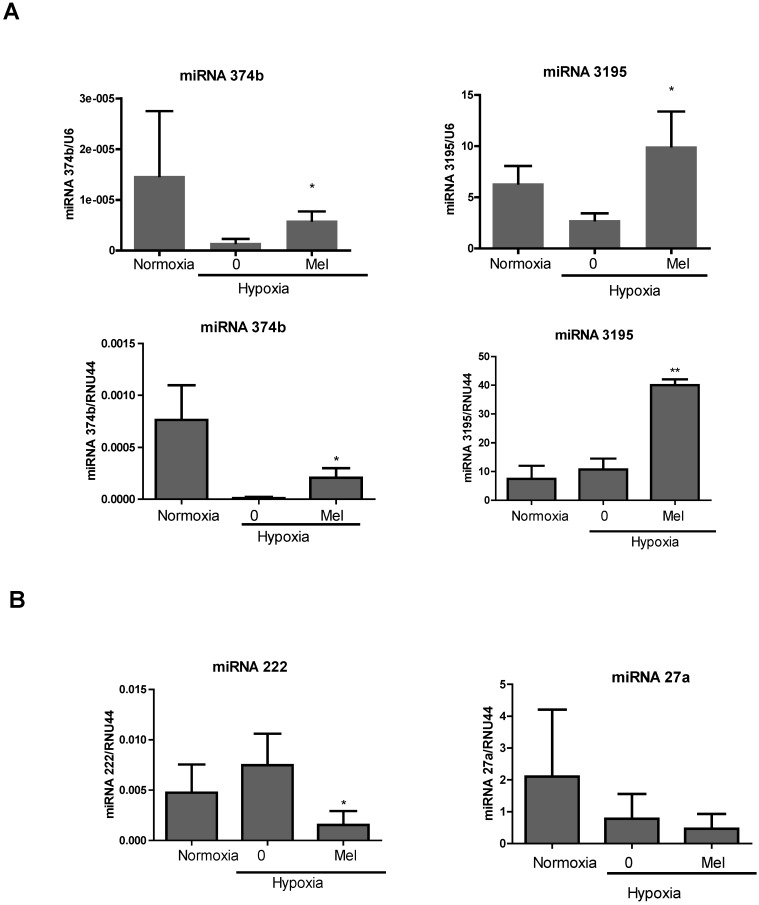
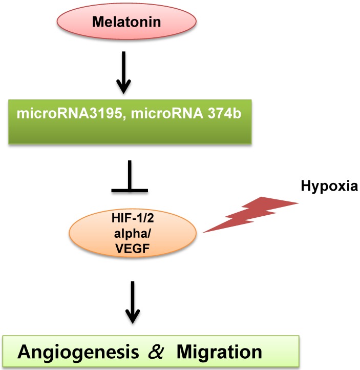
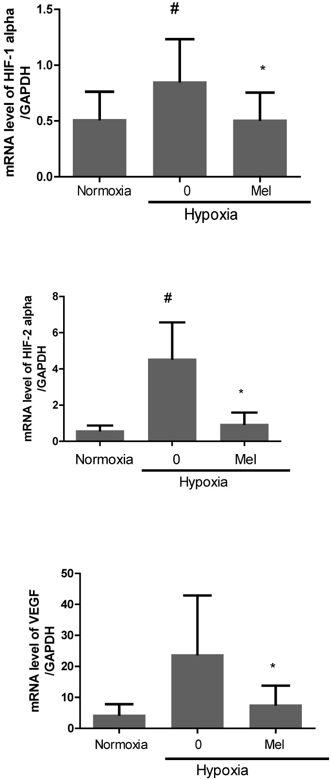
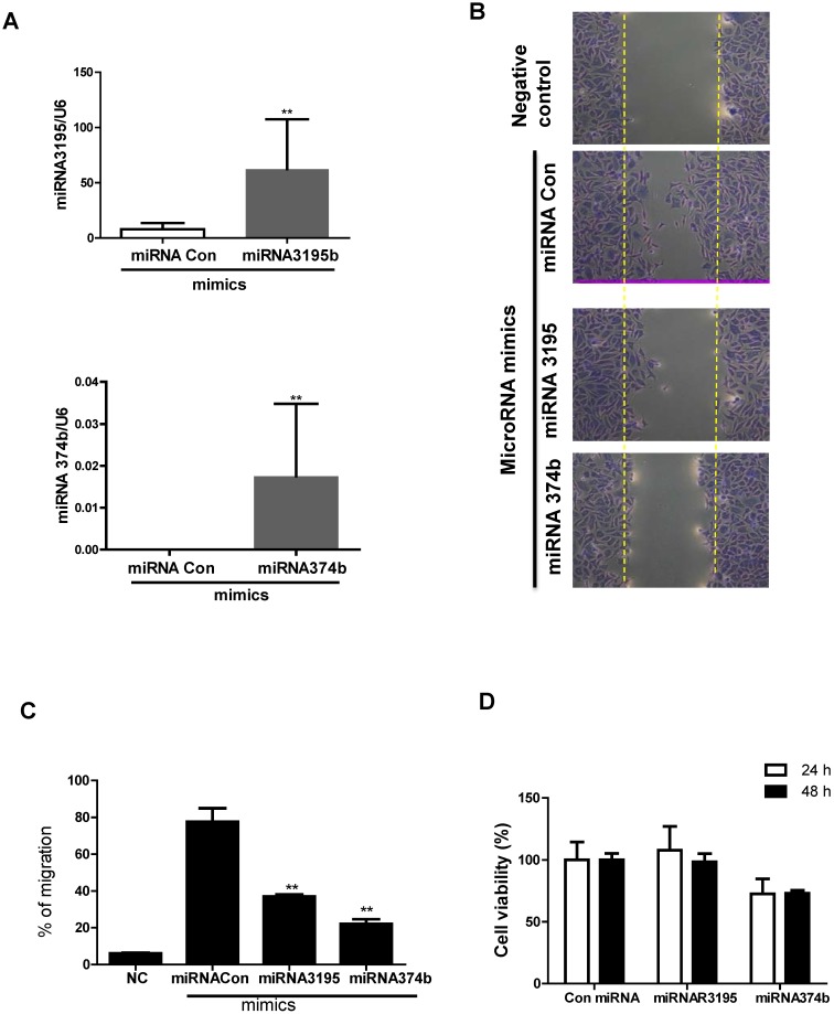
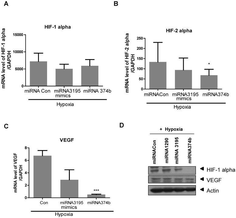
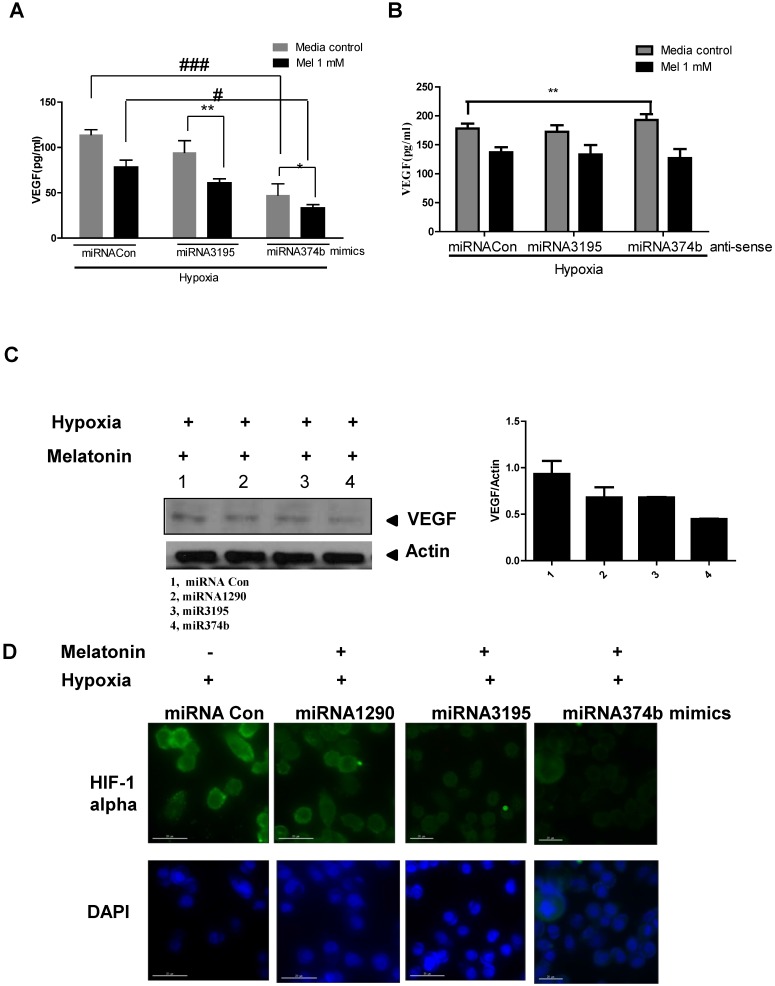
Recently microRNAs (miRNAs) have been attractive targets with their key roles in biological regulation through post-transcription to control mRNA stability and protein translation. Though melatonin was known as an anti-angiogenic agent, the underlying mechanism of melatonin in PC-3 prostate cancer cells under hypoxia still remains unclear. Thus, in the current study, we elucidated the important roles of miRNAs in melatonin-induced anti-angiogenic activity in hypoxic PC-3 cells. miRNA array revealed that 33 miRNAs (>2 folds) including miRNA3195 and miRNA 374b were significantly upregulated and 16 miRNAs were downregulated in melatonin-treated PC-3 cells under hypoxia compared to untreated control. Melatonin significantly attenuated the expression of hypoxia-inducible factor (HIF)-1 alpha, HIF-2 alpha and vascular endothelial growth factor (VEGF) at mRNA level in hypoxic PC-3 cells. Consistently, melatonin enhanced the expression of miRNA3195 and miRNA 374b in hypoxic PC-3 cells by qRT-PCR analysis. Of note, overexpression of miRNA3195 and miRNA374b mimics attenuated the mRNA levels of angiogenesis related genes such as HIF-1alpha, HIF-2 alpha and VEGF in PC-3 cells under hypoxia. Furthermore, overexpression of miRNA3195 and miRNA374b suppressed typical angiogenic protein VEGF at the protein level and VEGF production induced by melatonin, while antisense oligonucleotides against miRNA 3195 or miRNA 374b did not affect VEGF production induced by melatonin. Also, overexpression of miR3195 or miR374b reduced HIF-1 alpha immunofluorescent expression in hypoxic PC-3 compared to untreated control. Overall, our findings suggest that upregulation of miRNA3195 and miRNA374b mediates anti-angiogenic property induced by melatonin in hypoxic PC-3 cells.
最近,miRNAs(microRNAs)因其在通过转录后控制 mRNA 稳定性和蛋白质翻译来调节生物的关键作用而成为有吸引力的靶点。虽然褪黑素已被认为是一种抗血管生成剂,但褪黑素在低氧条件下对 PC-3 前列腺癌细胞的作用机制仍不清楚。因此,在本研究中,我们阐明了 miRNAs 在褪黑素诱导的低氧 PC-3 细胞抗血管生成活性中的重要作用。miRNA 芯片显示,与未经处理的对照组相比,在低氧条件下,褪黑素处理的 PC-3 细胞中有 33 种 miRNA(>2 倍)包括 miRNA3195 和 miRNA 374b 显著上调,而 16 种 miRNA 下调。褪黑素在低氧 PC-3 细胞中显著下调 HIF-1α、HIF-2α 和血管内皮生长因子(VEGF)的 mRNA 水平。一致地,褪黑素通过 qRT-PCR 分析增强了低氧 PC-3 细胞中 miRNA3195 和 miRNA 374b 的表达。值得注意的是,miRNA3195 和 miRNA374b 模拟物的过表达减弱了低氧条件下 PC-3 细胞中血管生成相关基因如 HIF-1α、HIF-2α 和 VEGF 的 mRNA 水平。此外,miRNA3195 和 miRNA374b 的过表达抑制了典型的血管生成蛋白 VEGF 的蛋白水平和褪黑素诱导的 VEGF 产生,而针对 miRNA3195 或 miRNA374b 的反义寡核苷酸则不影响褪黑素诱导的 VEGF 产生。同样,miR3195 或 miR374b 的过表达降低了低氧 PC-3 中 HIF-1α 的免疫荧光表达,与未经处理的对照组相比。总的来说,我们的研究结果表明,miRNA3195 和 miRNA374b 的上调介导了褪黑素在低氧 PC-3 细胞中的抗血管生成作用。