Biezonski Dominik K, Trifilieff Pierre, Meszaros Jozsef, Javitch Jonathan A, Kellendonk Christoph
Department of Psychiatry, Division of Molecular Therapeutics, Columbia University, New York State Psychiatric Institute, New York, New York, 10032, USA.
J Comp Neurol. 2015 Jun 1;523(8):1175-89. doi: 10.1002/cne.23730. Epub 2015 Feb 17.
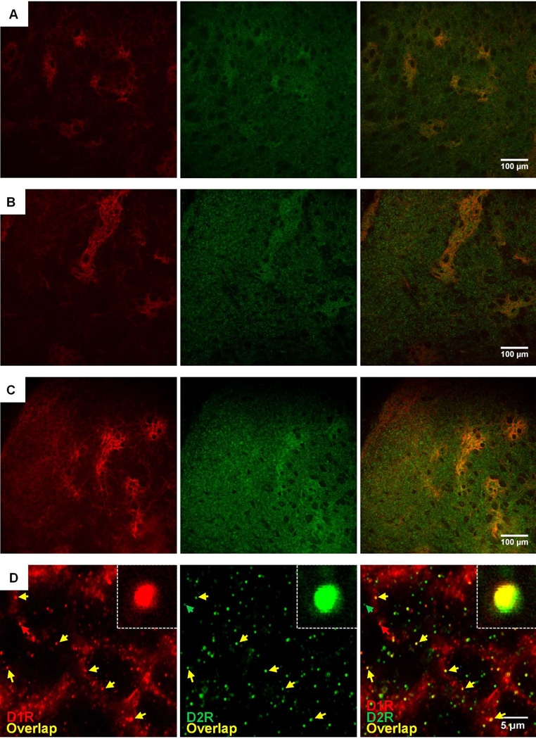
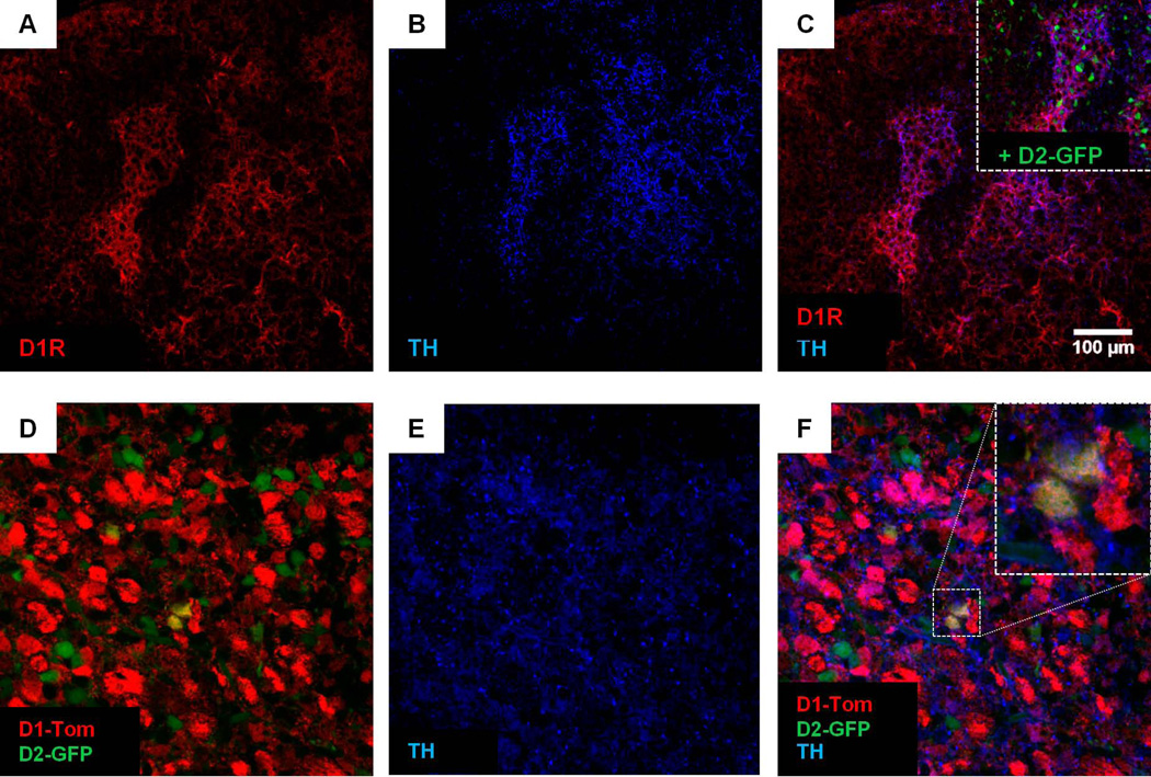
The striatum is the major input nucleus of the basal ganglia involved in reward processing, goal-directed behaviors, habit learning, and motor control. The striatum projects to the basal ganglia output nuclei via the "direct" and "indirect" pathways, which can be distinguished by their projection fields and their opposing effects on behavior. In adult animals, the functional opposition is modulated by the differential actions of D1 and D2 dopamine receptors (D1R, D2R), the expression of which is largely separated between these pathways. To determine whether a similar degree of separation exists earlier in development, we used dual-label immunohistochemistry to map dorsal-striatal D1R and D2R expression at the promoter level in postnatal day 0 (PD0) Drd1a-tdTomato/Drd2-GFP BAC transgenic mice, and at the receptor level by costaining for native D1R and D2R in wildtype (WT) PD0 animals. To assess for potential molecular interactions between D1R and D2R we also employed a recently developed proximity-ligation assay (PLA). Limited coexpression and colocalization of the D1R and D2R proteins was found in clusters of neurons endemic to the "patch" compartment as identified by costaining with tyrosine hydroxylase, but not outside these clusters. Moreover, in contrast to our recent findings where we failed to detect a D1R-D2R PLA signal in the adult striatum, in PD0 striatum we did identify a clear PLA signal for this pair of receptors. This colocalization at close proximity points to a possible role for D1R/D2R-mediated crosstalk in early striatal ontogeny.
纹状体是基底神经节的主要输入核,参与奖赏处理、目标导向行为、习惯学习和运动控制。纹状体通过“直接”和“间接”通路投射到基底神经节输出核,这两条通路可通过其投射区域以及对行为的相反作用来区分。在成年动物中,这种功能上的对立由D1和D2多巴胺受体(D1R、D2R)的不同作用调节,它们的表达在这些通路之间大体上是分开的。为了确定在发育早期是否存在类似程度的分离,我们使用双标记免疫组织化学法,在出生后第0天(PD0)的Drd1a-tdTomato/Drd2-GFP BAC转基因小鼠中,在启动子水平绘制背侧纹状体D1R和D2R的表达图谱,并在野生型(WT)PD0动物中通过对天然D1R和D2R进行共染色,在受体水平绘制其表达图谱。为了评估D1R和D2R之间潜在的分子相互作用,我们还采用了最近开发的邻近连接分析(PLA)。通过酪氨酸羟化酶共染色鉴定,在“斑块”区特有的神经元簇中发现了D1R和D2R蛋白的有限共表达和共定位,但在这些簇之外未发现。此外,与我们最近在成年纹状体中未检测到D1R-D2R PLA信号的发现相反,在PD0纹状体中,我们确实为这对受体鉴定出了清晰的PLA信号。这种紧密的共定位表明D1R/D2R介导的串扰在早期纹状体个体发生中可能发挥作用。