Tomkins Matthew Robert, Liao David Shiqi, Docoslis Aristides
Department of Chemical Engineering Queen's University, Kingston, ON K7L 3N6, Canada.
Sensors (Basel). 2015 Jan 8;15(1):1047-59. doi: 10.3390/s150101047.
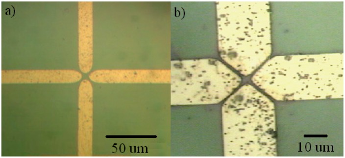
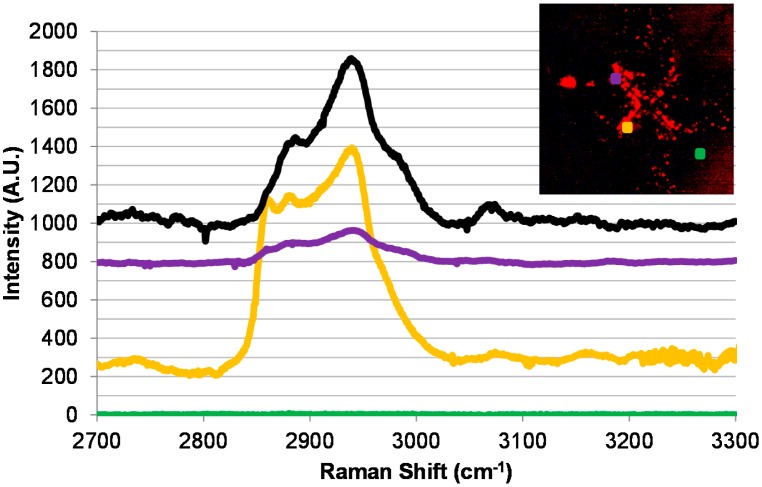
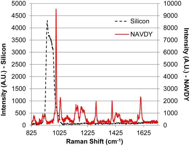
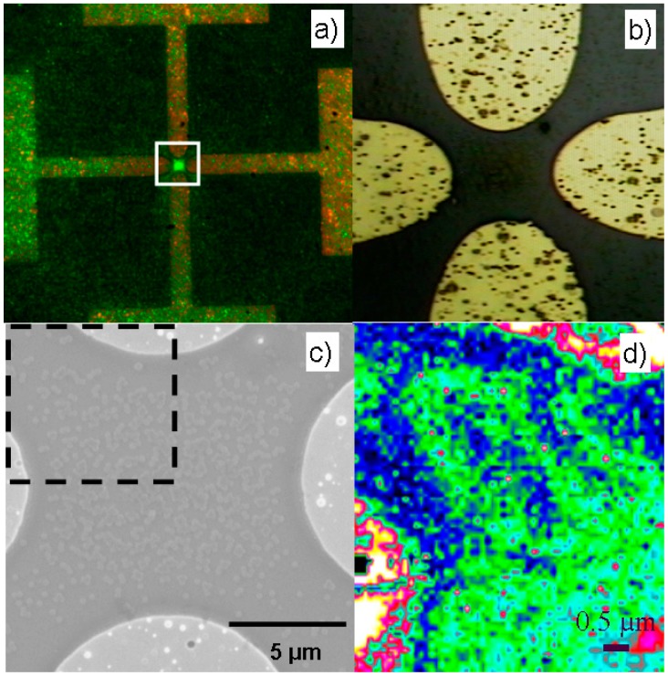
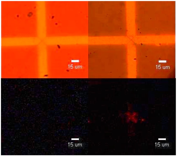
A detection method that combines electric field-assisted virus capture on antibody-decorated surfaces with the "fingerprinting" capabilities of micro-Raman spectroscopy is demonstrated for the case of M13 virus in water. The proof-of-principle surface mapping of model bioparticles (protein coated polystyrene spheres) captured by an AC electric field between planar microelectrodes is presented with a methodology for analyzing the resulting spectra by comparing relative peak intensities. The same principle is applied to dielectrophoretically captured M13 phage particles whose presence is indirectly confirmed with micro-Raman spectroscopy using NeutrAvidin-Cy3 as a labeling molecule. It is concluded that the combination of electrokinetically driven virus sampling and micro-Raman based signal transduction provides a promising approach for time-efficient and in situ detection of viruses.
本文展示了一种检测方法,该方法将抗体修饰表面上的电场辅助病毒捕获与显微拉曼光谱的“指纹识别”功能相结合,用于检测水中的M13病毒。通过平面微电极之间的交流电场捕获模型生物颗粒(蛋白质包被的聚苯乙烯球),给出了原理验证表面图谱,并介绍了一种通过比较相对峰强度来分析所得光谱的方法。同样的原理应用于介电泳捕获的M13噬菌体颗粒,使用中性抗生物素蛋白-Cy3作为标记分子,通过显微拉曼光谱间接确认其存在。得出的结论是,电动驱动的病毒采样与基于显微拉曼的信号转导相结合,为高效、原位检测病毒提供了一种有前景的方法。