Chan Siang-Hua Victor, Wong Kai-Sing Alain, Woo Yat-Ming Peter, Chan Kwong-Yau, Leung Kar-Ming
Department of Radiology, Queen Mary Hospital, Hong Kong S.A.R, China.
Department of Neurosurgery, Kwong Wah Hospital, Hong Kong S.A.R, China.
J Cerebrovasc Endovasc Neurosurg. 2014 Dec;16(4):358-63. doi: 10.7461/jcen.2014.16.4.358. Epub 2014 Dec 30.
Several modalities are available for volumetric measurement of the intracranial aneurysm. We discuss the challenges involved in manual segmentation, and analyze the application of alternative methods using automatic segmentation and geometric formulae in measurement of aneurysm volumes and coil packing density.
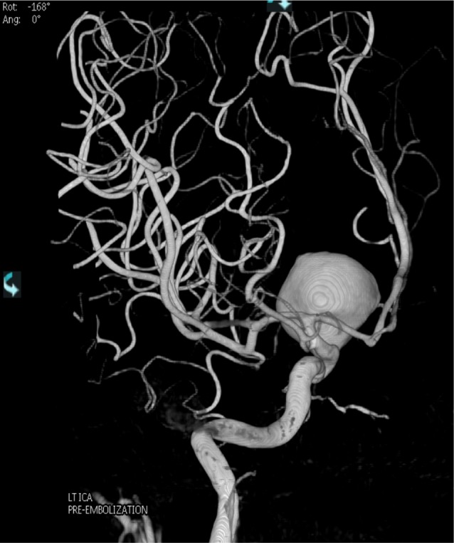
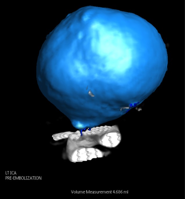
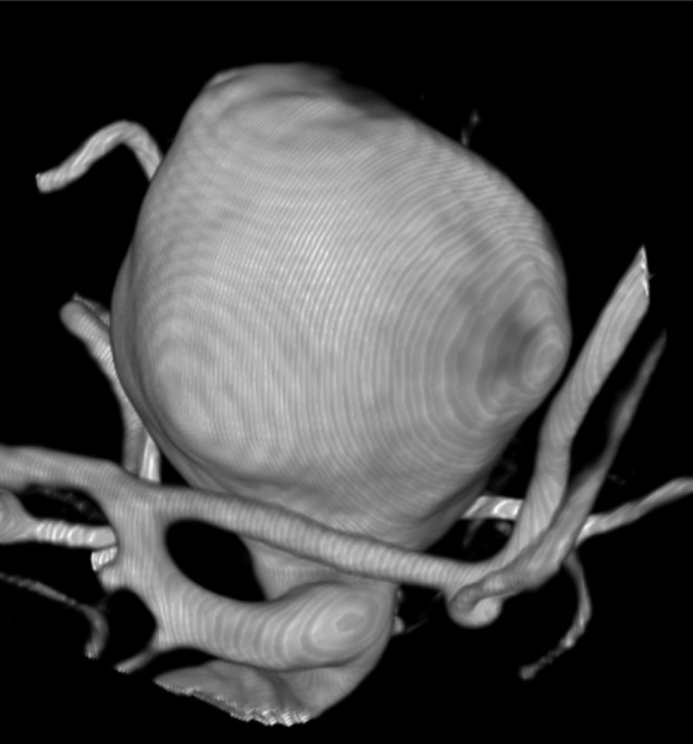
The volumes and morphology of 38 aneurysms treated with endovascular coiling at a single center were measured using three-dimensional rotational angiography (3DRA) reconstruction software using automatic segmentation. Aneurysm volumes were also calculated from their height, width, depth, size of neck, and assumed shape in 3DRA images using simple geometric formulae. The aneurysm volumes were dichotomized as "small" or "large" using the median volume of the studied population (54 mm(3)) measured by automatic segmentation as the cut-off value for further statistical analysis.
A greater proportion of aneurysms were categorized as being "small" when geometric formulae were applied. The median aneurysm volumes obtained were 54.5 mm(3) by 3DRA software, and 30.6 mm(3) using mathematical equations. An underestimation of aneurysm volume with a resultant overestimation in the calculated coil packing density (p = 0.002) was observed.
Caution must be exercised in the application of simple geometric formulae in the management of intracranial aneurysms as volumes may potentially be underestimated and packing densities falsely elevated. Future research should focus on validation of automatic segmentation in volumetric measurement and improving its accuracy to enhance its application in clinical practice.
有多种方法可用于颅内动脉瘤的体积测量。我们讨论了手动分割所涉及的挑战,并分析了使用自动分割和几何公式的替代方法在动脉瘤体积和线圈填充密度测量中的应用。
使用自动分割的三维旋转血管造影(3DRA)重建软件测量了在单一中心接受血管内栓塞治疗的38个动脉瘤的体积和形态。还使用简单的几何公式根据3DRA图像中的高度、宽度、深度、颈部大小和假定形状计算动脉瘤体积。以自动分割测量的研究人群中位数体积(54 mm³)作为进一步统计分析的临界值,将动脉瘤体积分为“小”或“大”。
应用几何公式时,更多比例的动脉瘤被归类为“小”。通过3DRA软件获得的动脉瘤中位数体积为54.5 mm³,使用数学公式为30.6 mm³。观察到动脉瘤体积被低估,导致计算的线圈填充密度高估(p = 0.002)。
在颅内动脉瘤的治疗中应用简单几何公式时必须谨慎,因为体积可能被低估,填充密度可能被错误提高。未来的研究应集中在体积测量中自动分割的验证以及提高其准确性,以增强其在临床实践中的应用。