• 文献检索
  • 文档翻译
  • 深度研究
  • 学术资讯
  • Suppr Zotero 插件Zotero 插件
  • 邀请有礼
  • 套餐&价格
  • 历史记录
应用&插件
Suppr Zotero 插件Zotero 插件浏览器插件Mac 客户端Windows 客户端微信小程序
定价
高级版会员购买积分包购买API积分包
服务
文献检索文档翻译深度研究API 文档MCP 服务
关于我们
关于 Suppr公司介绍联系我们用户协议隐私条款
关注我们

Suppr 超能文献

核心技术专利:CN118964589B侵权必究
粤ICP备2023148730 号-1Suppr @ 2026

文献检索

告别复杂PubMed语法,用中文像聊天一样搜索,搜遍4000万医学文献。AI智能推荐,让科研检索更轻松。

立即免费搜索

文件翻译

保留排版,准确专业,支持PDF/Word/PPT等文件格式,支持 12+语言互译。

免费翻译文档

深度研究

AI帮你快速写综述,25分钟生成高质量综述,智能提取关键信息,辅助科研写作。

立即免费体验

一种适用于任何形状和模态的动脉瘤体积量化的有效方法。

An Efficient Method for Aneurysm Volume Quantification Applicable in Any Shape and Modalities.

作者信息

Chung Jaewoo, Ko Jung Ho

机构信息

Department of Neurosurgery, Dankook University College of Medicine, Cheonan, Korea.

出版信息

J Korean Neurosurg Soc. 2021 Jul;64(4):514-523. doi: 10.3340/jkns.2020.0255. Epub 2021 Jun 29.

DOI:10.3340/jkns.2020.0255
PMID:34185985
原文链接:https://pmc.ncbi.nlm.nih.gov/articles/PMC8273770/
Abstract

OBJECTIVE

Aneurysm volume quantification (AVQ) using the equation of ellipsoid volume is widely used although it is inaccurate. Furthermore, AVQ with 3-dimensional (3D) rendered data has limitations in general use. A novel universal method for AVQ is introduced for any diagnostic modality and application to any shape of aneurysms.

METHODS

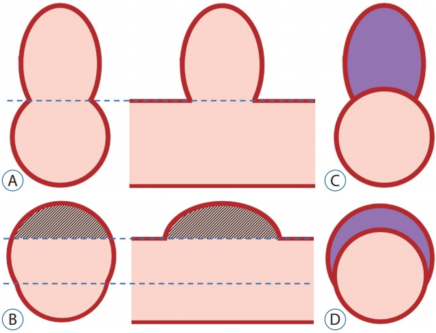
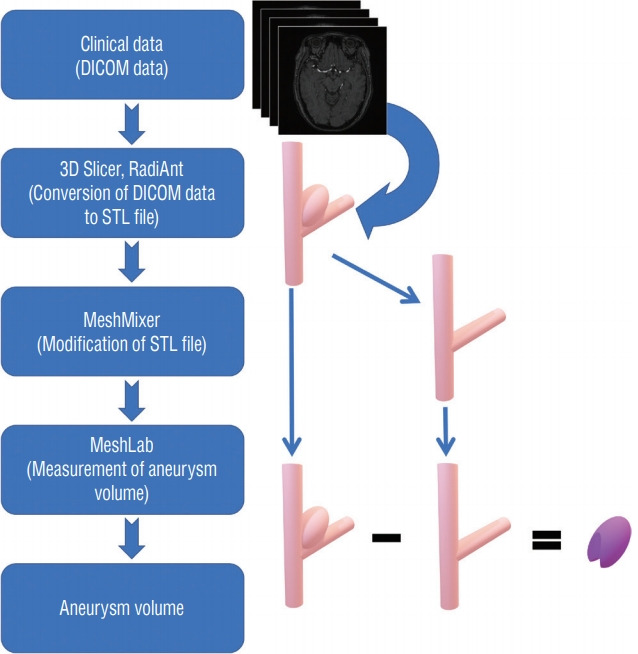
Relevant AVQ studies published from January 1997 to June 2019 were identified to determine common methods of AVQ. The basic idea is to eliminate the normal artery volume from 3D model with the aneurysm. After Digital Imaging and Communications in Medicine (DICOM) data is converted and exported to stereolithography (STL) file format, the 3D STL model is modified to remove the aneurysm and the volume difference between the 3D model with/without the aneurysm is defined as the aneurysm volume. Fifty randomly selected aneurysms from DICOM database were used to validate the different AVQ methods.

RESULTS

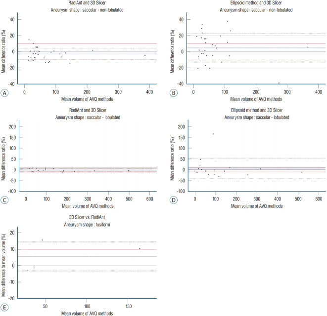
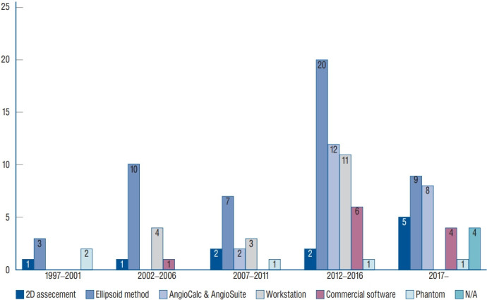
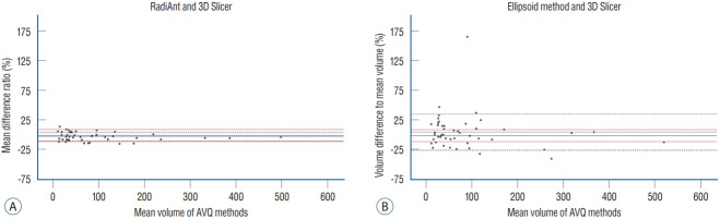
We reviewed and categorized AVQ methods in 121 studies. Approximately 60% used the ellipsoid method, while 24% used the 3D model. For 50 randomly selected aneurysms, volumes were measured using 3D Slicer, RadiAnt, and ellipsoid method. Using 3D Slicer as the reference, the ratios of mean difference to mean volume obtained by RadiAnt and ellipsoid method were -1.21±7.46% and 4.04±30.54%, respectively. The deviations between RadiAnt and 3D Slicer were small despite of aneurysm shapes, but those of ellipsoid method and 3D Slicer were large.

CONCLUSION

In spite of inaccuracy, ellipsoid method is still mostly used. We propose a novel universal method for AVQ that is valid, low cost, and easy to use.

摘要

目的

使用椭球体体积公式进行动脉瘤体积量化(AVQ)虽不准确但应用广泛。此外,基于三维(3D)渲染数据的AVQ在一般应用中存在局限性。本文介绍一种适用于任何诊断方式且可应用于任何形状动脉瘤的新型通用AVQ方法。

方法

检索1997年1月至2019年6月发表的相关AVQ研究,以确定常用的AVQ方法。基本思路是从包含动脉瘤的三维模型中去除正常动脉体积。将医学数字成像和通信(DICOM)数据转换并导出为立体光刻(STL)文件格式后,对三维STL模型进行修改以去除动脉瘤,将有/无动脉瘤的三维模型之间的体积差定义为动脉瘤体积。从DICOM数据库中随机选取50个动脉瘤用于验证不同的AVQ方法。

结果

我们对121项研究中的AVQ方法进行了综述和分类。约60%使用椭球体法,24%使用三维模型法。对于随机选取的50个动脉瘤,使用三维切片软件(3D Slicer)、RadiAnt和椭球体法测量体积。以3D Slicer为参照,RadiAnt法和椭球体法获得的平均差值与平均体积的比值分别为-1.21±7.46%和4.04±30.54%。尽管动脉瘤形状各异,但RadiAnt与3D Slicer之间的偏差较小,而椭球体法与3D Slicer之间的偏差较大。

结论

尽管不准确,但椭球体法仍被广泛使用。我们提出一种新型通用的AVQ方法,该方法有效、成本低且易于使用。

https://cdn.ncbi.nlm.nih.gov/pmc/blobs/b595/8273770/93d9bd0556b8/jkns-2020-0255f5.jpg
https://cdn.ncbi.nlm.nih.gov/pmc/blobs/b595/8273770/de757b10bfec/jkns-2020-0255f1.jpg
https://cdn.ncbi.nlm.nih.gov/pmc/blobs/b595/8273770/ded15207bd33/jkns-2020-0255f2.jpg
https://cdn.ncbi.nlm.nih.gov/pmc/blobs/b595/8273770/ca274b144015/jkns-2020-0255f3.jpg
https://cdn.ncbi.nlm.nih.gov/pmc/blobs/b595/8273770/da9290225cd8/jkns-2020-0255f4.jpg
https://cdn.ncbi.nlm.nih.gov/pmc/blobs/b595/8273770/93d9bd0556b8/jkns-2020-0255f5.jpg

相似文献

1
An Efficient Method for Aneurysm Volume Quantification Applicable in Any Shape and Modalities.一种适用于任何形状和模态的动脉瘤体积量化的有效方法。
J Korean Neurosurg Soc. 2021 Jul;64(4):514-523. doi: 10.3340/jkns.2020.0255. Epub 2021 Jun 29.
2
Accuracy of Voxel-Based and Algebraic Formula-Based Methods in Quantifying Cerebral Aneurysm Volume by 3D-Rotational Digital Subtraction Angiography. An In-Vitro and In-Vivo Study.基于体素法和代数公式法通过三维旋转数字减影血管造影术量化脑动脉瘤体积的准确性:一项体外和体内研究
Interv Neuroradiol. 2005 Mar 17;11(1):35-40. doi: 10.1177/159101990501100105. Epub 2005 Jun 17.
3
Stone volume measuring methods: Should the CT based three-dimensional-reconstructed algorithm be proposed as the gold standard? What did the three-dimensional printed models show us?结石体积测量方法:基于CT的三维重建算法应被视为金标准吗?三维打印模型向我们展示了什么?
Arch Esp Urol. 2019 Jul;72(6):596-601.
4
3D Printing of Rapid, Low-Cost and Patient-Specific Models of Brain Vasculature for Use in Preoperative Planning in Clipping of Intracranial Aneurysms.用于颅内动脉瘤夹闭术前规划的快速、低成本且个性化的脑血管模型的3D打印
J Clin Med. 2021 Mar 13;10(6):1201. doi: 10.3390/jcm10061201.
5
[Rapid 3-Dimensional Models of Cerebral Aneurysm for Emergency Surgical Clipping].[用于急诊手术夹闭的脑动脉瘤快速三维模型]
No Shinkei Geka. 2016 Aug;44(8):651-60. doi: 10.11477/mf.1436203350.
6
Quantitative three-dimensional reconstruction of aneurysmal left ventricles. In vitro and in vivo validation.左心室动脉瘤的定量三维重建。体外和体内验证。
Circulation. 1995 Jan 1;91(1):222-30. doi: 10.1161/01.cir.91.1.222.
7
Three-dimensional digital subtraction angiography vs two-dimensional digital subtraction angiography for detection of ruptured intracranial aneurysms: a study of 86 aneurysms.三维数字减影血管造影术与二维数字减影血管造影术用于检测破裂颅内动脉瘤的比较:86例动脉瘤的研究
Neurol India. 2005 Sep;53(3):287-9; discussion 290. doi: 10.4103/0028-3886.16925.
8
Dual-energy CT angiography in the evaluation of intracranial aneurysms: image quality, radiation dose, and comparison with 3D rotational digital subtraction angiography.双能量 CT 血管造影在颅内动脉瘤评估中的应用:图像质量、辐射剂量,以及与三维旋转数字减影血管造影的比较。
AJR Am J Roentgenol. 2010 Jan;194(1):23-30. doi: 10.2214/AJR.08.2290.
9
Usefulness of volume-rendered three-dimensional computed tomographic angiography for surgical planning in treating unruptured paraclinoid internal carotid artery aneurysms.容积再现三维计算机断层血管造影术在未破裂床突旁颈内动脉瘤手术规划中的应用价值
Kobe J Med Sci. 2001 Oct;47(5):221-30.
10
Accuracy and interobserver reliability of three-dimensional rotational angiography versus mathematical models for volumetric measurement of intracranial aneurysms.三维旋转血管造影术与数学模型用于颅内动脉瘤体积测量的准确性及观察者间可靠性比较
J Clin Neurosci. 2009 Sep;16(9):1195-8. doi: 10.1016/j.jocn.2008.11.011. Epub 2009 Jun 3.

引用本文的文献

1
A study on the therapeutic effect of precise clipping of intracranial aneurysms assisted by CTA and 3D-slicer software.一项关于CTA及3D-切片软件辅助下颅内动脉瘤精准夹闭治疗效果的研究
Front Surg. 2025 Feb 17;12:1535585. doi: 10.3389/fsurg.2025.1535585. eCollection 2025.
2
Volumetric surveillance of brain aneurysms: Pitfalls of MRA.脑动脉瘤的容积监测:MRA 的陷阱。
Interv Neuroradiol. 2023 Oct;29(5):532-539. doi: 10.1177/15910199221100619. Epub 2022 May 12.

本文引用的文献

1
SMART coils for intracranial aneurysm embolization: Follow-up outcomes.用于颅内动脉瘤栓塞的SMART线圈:随访结果。
J Clin Neurosci. 2019 Jan;59:93-97. doi: 10.1016/j.jocn.2018.10.132. Epub 2018 Nov 7.
2
Individualized reconstruction for severe periprosthetic fractures around the tumor prosthesis of knee under assistance of 3D printing technology: A case report.3D打印技术辅助下膝关节肿瘤假体周围严重假体周围骨折的个体化重建:1例报告
Medicine (Baltimore). 2018 Oct;97(42):e12726. doi: 10.1097/MD.0000000000012726.
3
New generation Hydrogel Endovascular Aneurysm Treatment Trial (HEAT): a study protocol for a multicenter randomized controlled trial.
新一代水凝胶血管内动脉瘤治疗试验(HEAT):一项多中心随机对照试验的研究方案
Neuroradiology. 2018 Oct;60(10):1075-1084. doi: 10.1007/s00234-018-2074-5. Epub 2018 Aug 17.
4
Comparative analysis of aneurysm volume by different methods based on angiography and computed tomography angiography.基于血管造影和计算机断层扫描血管造影的不同方法对动脉瘤体积的比较分析。
Neurosurg Rev. 2018 Oct;41(4):1013-1019. doi: 10.1007/s10143-018-0943-3. Epub 2018 Jan 16.
5
Tumor Volume Assessment in Low-Grade Gliomas: A Comparison of Preoperative Magnetic Resonance Imaging to Coregistered Intraoperative 3-Dimensional Ultrasound Recordings.低级别胶质瘤的肿瘤体积评估:术前磁共振成像与配准的术中三维超声记录的比较。
Neurosurgery. 2018 Aug 1;83(2):288-296. doi: 10.1093/neuros/nyx392.
6
Patient and aneurysm characteristics associated with rupture risk of multiple intracranial aneurysms in the anterior circulation system.前循环系统中与多发性颅内动脉瘤破裂风险相关的患者及动脉瘤特征。
Acta Neurochir (Wien). 2016 Jul;158(7):1367-75. doi: 10.1007/s00701-016-2826-0. Epub 2016 May 10.
7
Large Residual Volume, Not Low Packing Density, Is the Most Influential Risk Factor for Recanalization after Coil Embolization of Cerebral Aneurysms.大残余体积而非低填充密度是脑动脉瘤弹簧圈栓塞术后再通的最具影响力的危险因素。
PLoS One. 2016 May 6;11(5):e0155062. doi: 10.1371/journal.pone.0155062. eCollection 2016.
8
Validation of the Modified Raymond-Roy classification for intracranial aneurysms treated with coil embolization.经弹簧圈栓塞治疗的颅内动脉瘤改良Raymond-Roy分类法的验证
J Neurointerv Surg. 2016 Sep;8(9):927-33. doi: 10.1136/neurintsurg-2015-012035. Epub 2015 Oct 5.
9
Association Between Vascular Anatomy and Posterior Communicating Artery Aneurysms.血管解剖结构与后交通动脉瘤之间的关联
World Neurosurg. 2015 Nov;84(5):1251-5. doi: 10.1016/j.wneu.2015.05.078. Epub 2015 Jun 11.
10
Morphology Parameters for Mirror Posterior Communicating Artery Aneurysm Rupture Risk Assessment.用于评估镜像后交通动脉瘤破裂风险的形态学参数
Neurol Med Chir (Tokyo). 2015;55(6):498-504. doi: 10.2176/nmc.oa.2014-0390. Epub 2015 Jun 2.