• 文献检索
  • 文档翻译
  • 深度研究
  • 学术资讯
  • Suppr Zotero 插件Zotero 插件
  • 邀请有礼
  • 套餐&价格
  • 历史记录
应用&插件
Suppr Zotero 插件Zotero 插件浏览器插件Mac 客户端Windows 客户端微信小程序
定价
高级版会员购买积分包购买API积分包
服务
文献检索文档翻译深度研究API 文档MCP 服务
关于我们
关于 Suppr公司介绍联系我们用户协议隐私条款
关注我们

Suppr 超能文献

核心技术专利:CN118964589B侵权必究
粤ICP备2023148730 号-1Suppr @ 2026

文献检索

告别复杂PubMed语法,用中文像聊天一样搜索,搜遍4000万医学文献。AI智能推荐,让科研检索更轻松。

立即免费搜索

文件翻译

保留排版,准确专业,支持PDF/Word/PPT等文件格式,支持 12+语言互译。

免费翻译文档

深度研究

AI帮你快速写综述,25分钟生成高质量综述,智能提取关键信息,辅助科研写作。

立即免费体验

免疫功能正常患者颅内原发性中枢神经系统淋巴瘤的磁共振成像特征

MR imaging features of intracranial primary CNS lymphoma in immune competent patients.

作者信息

Mansour Asem, Qandeel Monther, Abdel-Razeq Hikmat, Abu Ali Hussain Ali

出版信息

Cancer Imaging. 2014 May 7;14(1):22. doi: 10.1186/1470-7330-14-22.

DOI:10.1186/1470-7330-14-22
PMID:25608570
原文链接:https://pmc.ncbi.nlm.nih.gov/articles/PMC4331827/
Abstract

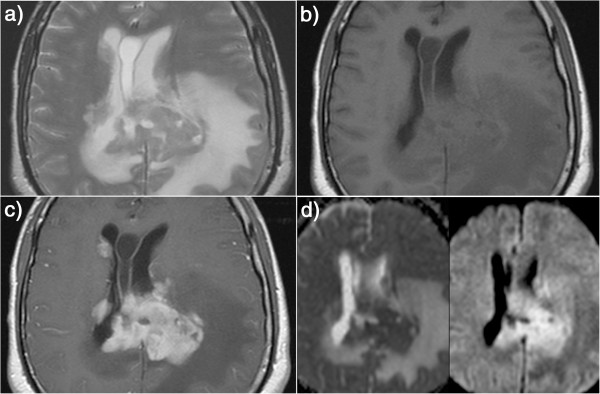
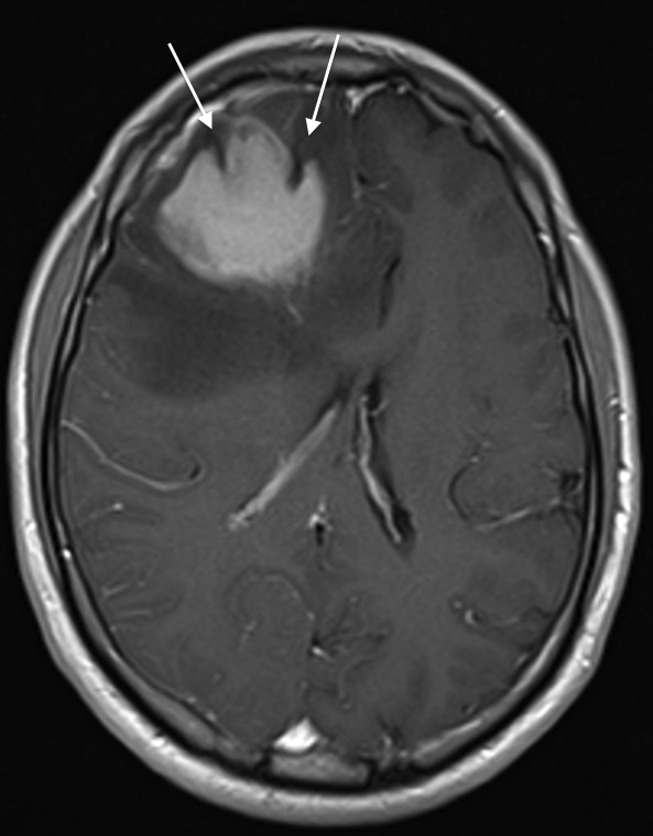
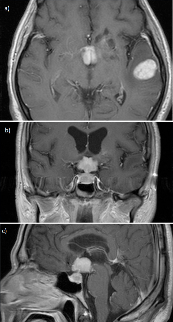
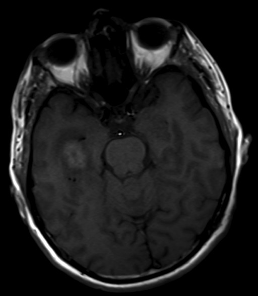
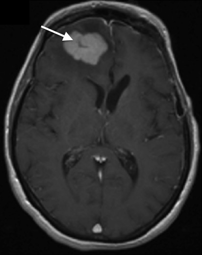
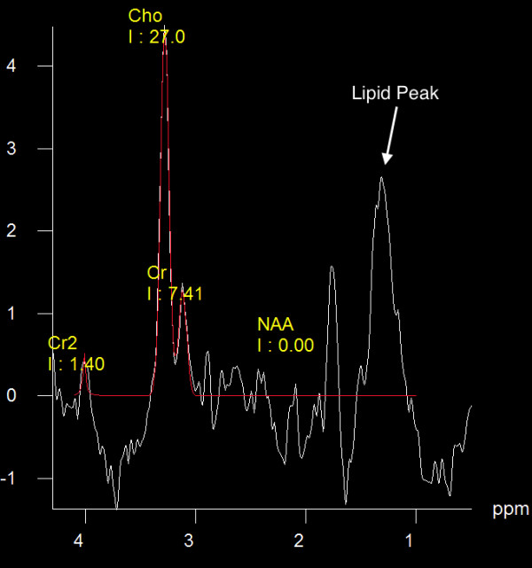
We aimed to characterize specific MRI findings seen in immune competent patients with intracranial primary CNS lymphoma (PCNSL) and to determine their value in the management of such patients. Pre-treatment MRI examinations of 21 immunocompetent patients with biopsy-proven PCNSL were retrospectively evaluated. T1 and T2 signal characteristics as well as contrast enhancement features are described in all patients. Diffusion, perfusion and proton-MR-spectroscopy features are described in a subset of these patients. In the proper clinical and radiologic setting, suggesting the diagnosis of PCNSL can help institute proper treatment in a timely fashion and avoid unnecessary attempts at surgical resection and the associated morbidity.

摘要

我们旨在描述免疫功能正常的颅内原发性中枢神经系统淋巴瘤(PCNSL)患者的特定MRI表现,并确定其在这类患者管理中的价值。对21例经活检证实为PCNSL的免疫功能正常患者的治疗前MRI检查进行了回顾性评估。描述了所有患者的T1和T2信号特征以及对比增强特征。在这些患者的一个子集中描述了扩散、灌注和质子磁共振波谱特征。在适当的临床和放射学背景下,提示PCNSL的诊断有助于及时进行适当的治疗,并避免不必要的手术切除尝试及相关并发症。

https://cdn.ncbi.nlm.nih.gov/pmc/blobs/e266/4331827/891db9647f91/1470-7330-14-22-7.jpg
https://cdn.ncbi.nlm.nih.gov/pmc/blobs/e266/4331827/89aeed820173/1470-7330-14-22-1.jpg
https://cdn.ncbi.nlm.nih.gov/pmc/blobs/e266/4331827/5b22d142119b/1470-7330-14-22-2.jpg
https://cdn.ncbi.nlm.nih.gov/pmc/blobs/e266/4331827/48aac9df44d8/1470-7330-14-22-3.jpg
https://cdn.ncbi.nlm.nih.gov/pmc/blobs/e266/4331827/5cb745101a76/1470-7330-14-22-4.jpg
https://cdn.ncbi.nlm.nih.gov/pmc/blobs/e266/4331827/1543e450caff/1470-7330-14-22-5.jpg
https://cdn.ncbi.nlm.nih.gov/pmc/blobs/e266/4331827/ea3b30075549/1470-7330-14-22-6.jpg
https://cdn.ncbi.nlm.nih.gov/pmc/blobs/e266/4331827/891db9647f91/1470-7330-14-22-7.jpg

相似文献

1
MR imaging features of intracranial primary CNS lymphoma in immune competent patients.免疫功能正常患者颅内原发性中枢神经系统淋巴瘤的磁共振成像特征
Cancer Imaging. 2014 May 7;14(1):22. doi: 10.1186/1470-7330-14-22.
2
Primary central nervous system lymphoma in immunocompetent patients: a retrospective review of MRI features.免疫功能正常患者的原发性中枢神经系统淋巴瘤:MRI特征的回顾性分析
J Med Imaging Radiat Oncol. 2012 Jun;56(3):295-301. doi: 10.1111/j.1754-9485.2012.02366.x. Epub 2012 Apr 10.
3
Primary central nervous system lymphoma and atypical glioblastoma: multiparametric differentiation by using diffusion-, perfusion-, and susceptibility-weighted MR imaging.原发性中枢神经系统淋巴瘤和非典型性脑胶质瘤:弥散、灌注和磁化率加权 MR 成像的多参数鉴别诊断。
Radiology. 2014 Sep;272(3):843-50. doi: 10.1148/radiol.14132740. Epub 2014 May 3.
4
Magnetic resonance features of primary central nervous system lymphoma in the immunocompetent patient: a pictorial essay.免疫功能正常患者原发性中枢神经系统淋巴瘤的磁共振成像特征:图文综述
J Med Imaging Radiat Oncol. 2012 Apr;56(2):179-86. doi: 10.1111/j.1754-9485.2012.02345.x. Epub 2012 Mar 6.
5
Radiomics features to distinguish glioblastoma from primary central nervous system lymphoma on multi-parametric MRI.基于多参数MRI的区分胶质母细胞瘤与原发性中枢神经系统淋巴瘤的影像组学特征
Neuroradiology. 2018 Dec;60(12):1297-1305. doi: 10.1007/s00234-018-2091-4. Epub 2018 Sep 19.
6
Central nervous system lymphoma characterization by diffusion-weighted imaging and MR spectroscopy.通过扩散加权成像和磁共振波谱对中枢神经系统淋巴瘤进行特征描述。
J Neuroimaging. 2008 Oct;18(4):411-7. doi: 10.1111/j.1552-6569.2007.00231.x. Epub 2008 May 19.
7
Inflammatory Demyelinating Lesions: True Sentinel Lesion or Immune-Mediated Response to Lymphoma?炎性脱髓鞘病变:真正的首发病变还是淋巴瘤的免疫介导反应?
World Neurosurg. 2021 Jan;145:172-177. doi: 10.1016/j.wneu.2020.09.064. Epub 2020 Sep 17.
8
Primary CNS lymphoma in Immunocompetent patients: Appearances on Conventional and Advanced Imaging with Review of literature.免疫功能正常的中枢神经系统原发性淋巴瘤:常规和先进影像学表现及文献复习。
J Radiol Case Rep. 2022 Jul 31;16(7):1-17. doi: 10.3941/jrcr.v16i7.4562. eCollection 2022 Jul.
9
Parenchymal lymphoma of the brain on initial MR imaging: a comparative study between primary and secondary brain lymphoma.脑实质淋巴瘤的初始磁共振成像表现:原发性与继发性脑淋巴瘤的对比研究。
Eur J Radiol. 2011 Aug;79(2):288-94. doi: 10.1016/j.ejrad.2010.01.017. Epub 2010 Mar 3.
10
Differentiation of glioblastoma and primary CNS lymphomas using susceptibility weighted imaging.应用磁敏感加权成像鉴别脑胶质母细胞瘤和原发性中枢神经系统淋巴瘤。
Eur J Radiol. 2013 Mar;82(3):552-6. doi: 10.1016/j.ejrad.2012.11.002. Epub 2012 Dec 10.

引用本文的文献

1
CNS Non-Hodgkin's Lymphoma With Atypical T2 Hyperintensity and Necrosis Mimicking Glioblastoma Multiforme: A Radiological Enigma.具有非典型T2高信号和坏死、酷似多形性胶质母细胞瘤的中枢神经系统非霍奇金淋巴瘤:一个影像学谜团。
Cureus. 2025 Aug 4;17(8):e89353. doi: 10.7759/cureus.89353. eCollection 2025 Aug.
2
Qualitative MRI features in the differentiation between primary and secondary CNS lymphoma.原发性与继发性中枢神经系统淋巴瘤鉴别中的磁共振成像定性特征
Neuroradiology. 2025 Mar 17. doi: 10.1007/s00234-025-03594-1.
3
Magnetic resonance spectroscopy for discriminating primary angiitis of the central nervous system from gliomas and lymphomas.

本文引用的文献

1
Neuroimaging of primary central nervous system lymphoma in immunocompetent patients: comparison of recent and previous findings.免疫功能正常患者原发性中枢神经系统淋巴瘤的神经影像学:近期与既往研究结果比较
J Nippon Med Sch. 2013;80(3):174-83. doi: 10.1272/jnms.80.174.
2
Cyst with a mural nodule tumor of the brain.脑内壁结节肿瘤性囊肿。
Cancer Imaging. 2012 Aug 10;12(1):237-44. doi: 10.1102/1470-7330.2012.0028.
3
Central nervous system lymphoma: characteristic findings on traditional and advanced imaging.中枢神经系统淋巴瘤:传统和先进成像技术的特征表现。
用于鉴别中枢神经系统原发性血管炎与胶质瘤和淋巴瘤的磁共振波谱分析。
Acta Neurol Belg. 2025 Apr;125(2):509-517. doi: 10.1007/s13760-025-02744-9. Epub 2025 Feb 11.
4
Utilizing machine-learning techniques on MRI radiomics to identify primary tumors in brain metastases.利用磁共振成像(MRI)影像组学的机器学习技术识别脑转移瘤中的原发肿瘤。
Front Neurol. 2025 Jan 6;15:1474461. doi: 10.3389/fneur.2024.1474461. eCollection 2024.
5
Isolated Third Cranial Nerve Palsy As the Initial Presentation of Primary Central Nervous System Lymphoma: A Rare Case.孤立性第三脑神经麻痹作为原发性中枢神经系统淋巴瘤的首发表现:1例罕见病例
Cureus. 2024 Dec 14;16(12):e75715. doi: 10.7759/cureus.75715. eCollection 2024 Dec.
6
[Primary central nervous system lymphoma in immunocompetent patient: A case report].[免疫功能正常患者的原发性中枢神经系统淋巴瘤:一例报告]
Rev Med Inst Mex Seguro Soc. 2024 Jul 1;62(4):1-6. doi: 10.5281/zenodo.11397215.
7
Mind Games: A Curious Case of Primary CNS Lymphoma Masquerading as Glioblastoma Multiforme.思维游戏:一例罕见的原发性中枢神经系统淋巴瘤伪装成多形性胶质母细胞瘤的病例
Cureus. 2024 Oct 13;16(10):e71358. doi: 10.7759/cureus.71358. eCollection 2024 Oct.
8
Differentiating between Lymphoma and Metastasis Presenting as Solid Cerebellar Mass Lacking Necrosis.鉴别表现为无坏死的实性小脑肿块的淋巴瘤与转移瘤。
Diagnostics (Basel). 2024 Oct 6;14(19):2228. doi: 10.3390/diagnostics14192228.
9
Primary central nervous system lymphomas in immunocompromised patients require specific response criteria.原发性中枢神经系统淋巴瘤在免疫功能低下的患者中需要特定的反应标准。
J Neurooncol. 2024 Aug;169(1):51-60. doi: 10.1007/s11060-024-04694-3. Epub 2024 Jun 12.
10
Primary central nervous system lymphoma: Imaging features and differential diagnosis.原发性中枢神经系统淋巴瘤:影像特征与鉴别诊断。
Neuroradiol J. 2024 Dec;37(6):705-722. doi: 10.1177/19714009241252625. Epub 2024 May 4.
AJNR Am J Neuroradiol. 2011 Jun-Jul;32(6):984-92. doi: 10.3174/ajnr.A2171. Epub 2010 Jul 8.
4
MRI findings in lymphomatosis cerebri: description of a case and revision of the literature.脑淋巴瘤的 MRI 表现:一例病例描述并文献复习。
J Neuroimaging. 2011 Apr;21(2):e183-6. doi: 10.1111/j.1552-6569.2010.00477.x.
5
Diffusion-weighted MR imaging derived apparent diffusion coefficient is predictive of clinical outcome in primary central nervous system lymphoma.弥散加权磁共振成像衍生的表观扩散系数可预测原发性中枢神经系统淋巴瘤的临床结局。
AJNR Am J Neuroradiol. 2010 Jan;31(1):60-6. doi: 10.3174/ajnr.A1750. Epub 2009 Sep 3.
6
CT and MR imaging features of primary central nervous system lymphoma in Norway, 1989-2003.1989 - 2003年挪威原发性中枢神经系统淋巴瘤的CT和MR成像特征
AJNR Am J Neuroradiol. 2009 Apr;30(4):744-51. doi: 10.3174/ajnr.A1447. Epub 2009 Jan 22.
7
Central nervous system lymphoma characterization by diffusion-weighted imaging and MR spectroscopy.通过扩散加权成像和磁共振波谱对中枢神经系统淋巴瘤进行特征描述。
J Neuroimaging. 2008 Oct;18(4):411-7. doi: 10.1111/j.1552-6569.2007.00231.x. Epub 2008 May 19.
8
Adjacent epidermoid cyst and primary central nervous system lymphoma: case report.相邻表皮样囊肿与原发性中枢神经系统淋巴瘤:病例报告
Surg Neurol. 2008 May;69(5):530-3; discussion 533-4. doi: 10.1016/j.surneu.2007.02.057. Epub 2008 Feb 8.
9
Primary central nervous system lymphoma.原发性中枢神经系统淋巴瘤
Neurol Clin. 2007 Nov;25(4):1193-207, xi. doi: 10.1016/j.ncl.2007.07.001.
10
Brain lymphoma: usefulness of the magnetic resonance spectroscopy.脑淋巴瘤:磁共振波谱分析的应用价值
J Neurooncol. 2008 Jan;86(2):225-9. doi: 10.1007/s11060-007-9468-2. Epub 2007 Sep 4.